241 posters, 10 topics, 7 sessions, 899 authors, 296 institutions
ePostersLive by SciGen Technologies S.A. All rights reserved.
The AAPS 103rd Annual Meeting 2025
May 24-27, 2025 | Austin, Texas

P15
How Flow Dynamics Influence Angiogenic Factor Release In Endothelial Cells Undergoing Micropuncture
Part of Topic
Basic Science Research
How Flow Dynamics Influence Angiogenic Factor Release in Endothelial Cells Undergoing Micropuncture
Mohammad Hossein Asgardoon, MD, MPH, Shailaja Agrawal, PhD, Mary Landmesser, BS, Maryam Abdelaal, BS, Jazzmyn Dawes, BS, Dino Ravnic, DO, MPH
Pennsylvania State University, College of Medicine, Hershey, PA, USA
Introduction
•Poor vascularization is a critical factor limiting graft survival in reconstructive surgery.
•Micropuncture (MP) is a novel surgical technique that can be used to enhance angiogenesis out of a targeted macrovasculature1.
•In MP, we use a 60 µm-diameter needle (Figure 1A) to precisely perforate the blood vessel wall at defined intervals to stimulate microvascular growth (Figure 1B).
•Static in vitro testing suggests that venous MP expedites microvascular formation compared to arterial MP via angiogenic factor release but the significance of flow to the process is unknown.
•We hypothesize that flow further augments angiogenic factor secretion following MP from both arterial and venous endothelial cells (ECs).
Methods
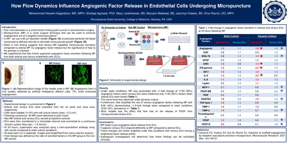
•Experimental design is summarized in Figure 2.
•Arterial and venous ECs were extracted from the rat aorta and vena cava, respectively.
•Cells were seeded (1x10⁵) onto cell culture inserts (area = 4.2 cm²).
•Following confluence, 40 MPs were delivered to each insert.
•Non-MP arterial and venous ECs served as baseline controls.
•ECs were then transferred to a microslide channel and connected to a continuous infusion system (flow rate = 1.5 ml/min).
•At 24 hours, supernatant was analyzed using a semi-quantitative antibody array, with results compared to static culture conditions.
•All tests were run in duplicate. ImageJ and GraphPad Prism were used for analysis.
•Fold change was defined as the ratio of secreted factors in the MP group to the non-MP control.
Results
•Under static conditions, MP was associated with >1-fold change of 17/20 (85%) angiogenic factors when venous ECs were tested but only 11/20 (55%) factors when arterial ECs were tested (Table 1).
•The same trend was observed under dynamic culture.
•Furthermore, flow amplified the rise of venous angiogenic factors following MP with 8/20 (40%) demonstrating >1.5-fold change when compared to static conditions (5/20, 25%, denoted by ; Table 1)
•Most notable was the effect that flow had on the release of PDGF from micropunctured venous ECs.
Conclusions
•MP leads to pro-angiogenic factor release from ECs.
•It appears venous ECs respond differently to MP compared to arterial ECs.
•These changes are further amplified under flow conditions with venous ECs having a heightened factor release profile.
•Subsequent investigations will determine how these findings can be translated clinically.