203 posters, 14 topics, 5 sessions, 817 authors, 256 institutions
ePostersLive by SciGen Technologies S.A. All rights reserved.
The AAPS 102nd Annual Meeting
May 18-21, 2024 | The Westin Copley Place, Boston, MA

D137
Artificial Intelligence Based Classification Of ICG Lymphography Patterns
Poster Presenter
Part of Topic
Education/Practice Management/Other
Artificial Intelligence Based Classification of ICG Lymphography Patterns
Berk B Ozmen, MD, Sonia K Pandey, MD, Graham S Schwarz, MD, MSE, FACS
Department of Plastic Surgery, Cleveland Clinic
Background
Indocyanine green (ICG) lymphography is a valuable tool in diagnosing and managing lymphedema [1]. There are 5 established ICG lymphography patterns; linear, reticular, splash, stardust and diffuse [2]. Interpretation of ICG lymphography is subjective and relies on the clinician's expertise [3]. Artificial Intelligence (AI), particularly computer vision and convolutional neural networks (CNNs), could improve ICG lymphography analysis by automating and standardizing the interpretation process [4]. In this study, we aim to develop an AI model, specifically a CNN algorithm, in predicting and classifying ICG lymphography patterns; linear, reticular, splash, stardust and diffuse.
Methods
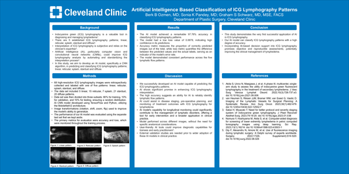
68 high-resolution ICG lymphography images were retrospectively collected and labeled with one of five patterns: linear, reticular, splash, stardust, and diffuse. The data set included 5 linear, 15 reticular, 7 splash, 21 stardust, 20 diffuse patterns. Data set was then divided into three subsets: 80% for training, 10% for validation, and 10% for testing, ensuring a random distribution. AI CNN model developed using TensorFlow and Python, utilizing the MobileNetV2 architecture. Image transformations (rotation, shift, zoom, flip) used to improve the model's ability to generalize. The performance of our AI model was evaluated using the separate test set that we kept aside. The primary metrics for evaluation were accuracy and loss, which were monitored throughout the training process.
Results
The AI model achieved a remarkable 97.78% accuracy in classifying ICG lymphography patterns. The model had a low loss value of 0.0678, indicating high confidence in its predictions. Accuracy metric measures the proportion of correctly predicted images out of the total, while loss metric quantifies the difference between the predicted values and the actual labels, serving as an indicator of the model's error rate. The model demonstrated consistent performance across the five lymphatic flow patterns.
Discussion
We successfully developed an AI model capable of predicting the ICG lymphogprahy patterns. AI shows significant promise in enhancing ICG lymphography interpretation. The high accuracy suggests an ability for AI to reliably identify lymphatic flow patterns. AI could assist in disease staging, pre-operative planning, and monitoring of treatment outcomes with ICG lymphography for lymphedema. AI model's capability for longitudinal monitoring could significantly contribute to the management of lymphatic disorders, offering a tool for early intervention and a broader application in clinical practice. Model performed across different images, without the need for specific anatomical considerations. User-friendly AI tools could improve diagnostic capabilities for trainees and early practitioners [5]. External validation studies are needed prior to wider adoption of these AI models in clinical practice.
Conclusion
This study demonstrates the very first successful application of AI in ICG lymphography. Our AI model can classify ICG lymphography patterns with a high degree of accuracy. Incorporating AI-based decision support into ICG lymphography promises objective and reproducible assessments, potentially improving the clinical management of lymphedema.
References
1.Akita S, Unno N, Maegawa J, et al. A phase III, multicenter, single-arm study to assess the utility of indocyanine green fluorescent lymphography in the treatment of secondary lymphedema. J Vasc Surg Venous Lymphat Disord. 2022;10(3):728-737.e3. doi:10.1016/j.jvsv.2021.09.006
2. van Heumen S, Riksen JJM, Bramer WM, van Soest G, Vasilic D. Imaging of the Lymphatic Vessels for Surgical Planning: A Systematic Review. Ann Surg Oncol. 2023;30(1):462-479. doi:10.1245/s10434-022-12552-7
3. Sakai H, Miyazaki T. Need for better protocol and severity staging system of indocyanine green lymphography. J Plast Reconstr Aesthet Surg. 2023;79:19-20. doi:10.1016/j.bjps.2023.01.038
4. Nomura Y, Hoshiyama M, Akita S, et al. Computer-aided diagnosis for screening of lower extremity lymphedema in pelvic computed tomography images using deep learning. Sci Rep. 2023;13(1):16214. doi:10.1038/s41598-023-43503-1
5. Dip F, Alexandru N, Amore M, et al. Use of fluorescence imaging during lymphatic surgery: A Delphi survey of experts worldwide. Surgery. 2022;172(6, Supplement):S14-S20. doi:10.1016/j.surg.2022.08.026