171 posters, 10 topics, 5 sessions, 690 authors, 211 institutions
ePostersLive by SciGen Technologies S.A. All rights reserved.
American Association of Plastic Surgeons Meeting 2022
April 9 - 12 | San Diego, California

P33
Peg-fusion To Restore Electrophysiological Continuity And Function After Nerve Transection
Poster Presenter
Authors
Affiliations
Part of Topic
Basic Science Research
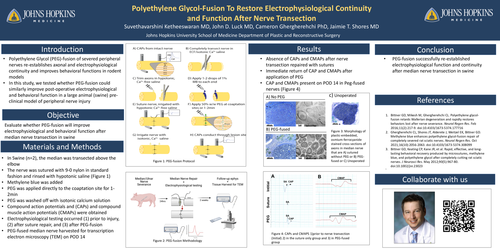
Background: Polyethylene Glycol (PEG)-fusion of severed peripheral nerves has been shown to re-establish axonal and electrophysiological continuity, and improve behavioral functions in rodent models. In this study, we tested whether PEG-fusion could similarly improve post-operative electrophysiological and behavioral functions in a large animal (swine) pre-clinical model of peripheral nerve injury.
Method: Yorkshire pigs (n=2) were used in a median/ulnar nerve transection model. The median and ulnar nerves were both sharply transected above the elbow. The ulnar nerve was left unrepaired, while the median nerve was repaired with standard suture repair followed by PEG-fusion. Immediately after transection, the median nerve was irrigated with hypotonic calcium-free saline followed by application of 1-2 drops of methylene blue to the transected endings. The nerve was sutured with 9-0 nylon in standard fashion. PEG was applied directly to the coaptation site for 1-2min. After 1-2min, PEG was washed off with isotonic Lactated Ringers solution. Electrophysiological testing of compound action potentials (CAPs) and compound muscle action potentials (CMAPs) was performed to measure if PEG resulted in the re-establishment of electrical connectivity between proximal and distal nerve segments. Stimulating electrodes were placed on the proximal end of the median nerve, while recording electrodes were placed on the nerve (CAPs) or in the flexor muscles of the anterior forearm muscles (CMAPs). Electrophysiological testing occurred (1) prior to injury, (2) after suture repair, and (3) after PEG-fusion.
Results Nerve conduction testing demonstrated the absence of CAPs or CMAPs after the nerve was repaired with sutures, but the immediate return of CAP and CMAPs after application of PEG. Follow up nerve conduction studies performed 14 days post-operatively showed continual maintenance of CAPs in both pigs, and CMAPs in 1 of 2 pigs, suggesting axonal survival and maintenance of some endplate innervation.
Conclusion PEG-fusion successfully re-established electrophysiological function and continuity after median nerve transection in swine. These results suggest that PEG-fusion of peripheral nerves is possible not only in rodents, but also in larger mammalian animal models. Additional studies in swine will be useful in bridging PEG-fusion from rodents to clinical studies. Swine could be a particularly useful model for PEG-fusion of long nerve defects (>3cm) that are not possible in rodents or other small animal models.