171 posters, 10 topics, 5 sessions, 690 authors, 211 institutions
ePostersLive by SciGen Technologies S.A. All rights reserved.
American Association of Plastic Surgeons Meeting 2022
April 9 - 12 | San Diego, California

PC2
Three-dimensional Tissue Rendering And Volume Analysis Of A Novel Medial Thigh Flap Using CT Angiography: The Chimeric Gracilis And PAP Flap
Poster Presenter
Authors
Jacob Hammond, Lyndsay Kandi, Chad Teven, Clint Jokerst, Jonathan Flug, Max Shrout, Marko Laitinen, Erwin Kruger, William Casey III, Alanna Rebecca
Affiliations
Part of Topic
Basic Science Research
Three-dimensional Tissue Rendering And Volume Analysis Of A Novel Medial Thigh Flap Using CT Angiography: The Chimeric Gracilis And PAP Flap
Jacob B. Hammond MD, Lyndsay Kandi BS, Chad M. Teven MD, Clint E. Jokerst MD, Jonathan A. Flug MD, Max A. Shrout MD, Marko A.Laitinen MD, Erwin A. Kruger MD, William J. Casey III MD, Alanna
Background
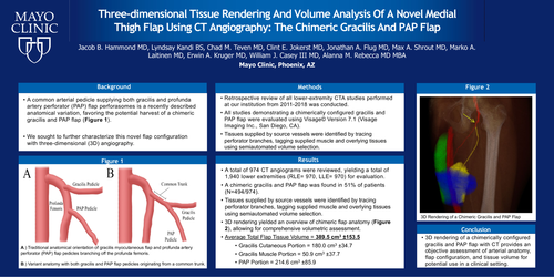
A common arterial pedicle supplying both gracilis and profunda artery perforator (PAP) flap perforasomes is a recently described anatomical variation, favoring the potential harvest of a chimeric gracilis and PAP flap. We sought to further characterize this novel flap configuration with three-dimensional (3D) angiography.
Methods
Lower extremity CT angiograms were performed according to standard angiography technique: 5 ml/s contrast injection with bolus tracking. Thin slice (0.6mm) axial reconstructions created multiplanar and volume rendered 3D images. Post-processing (Visage 7.1.15b) consisting of vessel measurements and centerline reconstructions of gracilis and PAP pedicles. Tissues supplied by these vessels were identified by tracing perforator branches, tagging supplied muscle and overlying tissues using semiautomated volume selection.
Results
A total of 974 CT angiograms were reviewed, yielding a total of 1,940 lower extremities (RLE= 970, LLE= 970) for evaluation. A chimeric gracilis and PAP flap was found in 51% of patients (N=494/974). Average length of the common arterial pedicle was 31.1 ±16.5 mm (Range = 2.0-95 mm), with an average diameter of 2.8 ±0.7 mm (Range = 1.3-8.8 mm). Average length of downstream gracilis and PAP flap pedicles were 58.0 ±18.7 mm (Range = 9.8-123 mm) and 101.7±27.0 mm (Range = 42.2-183 mm) respectively. 3D rendering yielded an overview of chimeric flap anatomy (Figure 1), allowing for comprehensive volumetric assessment (197.1 ± 29.2 cm3)
Conclusion
3D rendering of a chimerically configured gracilis and PAP flap with CT provides an objective assessment of arterial anatomy, flap configuration, and tissue volume for potential use in a clinical setting.