4,068 posters, 219 videos, 101 topics, 69 sessions, 14,365 authors, 6,128 institutions
ePostersLive by SciGen Technologies S.A. All rights reserved.
Alzheimer's Association International Conference 2026
July 12-15, 2026 | London, United Kingdom, and Online

13236
Mason Pirner, B.S.
Poster Presenter
Part of Topic
Basic Science and Pathogenesis -> Human Neuropathology -> Amyloid
Distinct Microglia-Plaque Spatial Relationships in Individuals Resilient to Alzheimer’s Disease
Mason B. Pirner1 , Nicholas E. Karagas1 , Corbin S. C. Johnson1 , Alexandra N. Cochoit1 , Isabel C. Smith1 , Rachel Blaine1 , Vanessa K. Souders1, C. Dirk Keene1, Thomas J. Grabowski1 , Caitlin S. Latimer1 , Kevin Z. Lin1 , Suman Jayadev1 , and Katherine E. Prater1 University of Washington. Seattle, WA 981051 Department of Neurology, University of Washington, Seattle, WA
Abstract / Background and Significance
Hypothesis: Cognitively resilient individuals with Alzheimer’s pathology will exhibit distinct microglia-Aβ colocalization patterns compared to symptomatic individuals
•Some older adults have Alzheimer’s pathology but remain cognitively intact—a phenomenon called resilience1 .
•These resilient individuals may have unique neuroimmune responses, particularly involving microglia2 .
•Microglia cluster around amyloid beta plaques, but their role in cognitive resilience is not yet well understood3 .
Methods
-
15µm-thick dorsolateral prefrontal cortical slices were stained for Iba1⁺ microglia and Pan-Aβ⁺ plaques
-
Immunofluorescence mosaics were acquired via stitching of confocal microscopy images
-
Surfaces characterizing the shape and area of microglia and plaques were generated using machine learning-based segmentation in IMARIS
-
Spatial overlap and proximity metrics were extracted, Spatial analysis was performed in Python to compare microglia-Aβ colocalization across groups
Results
Results: Microglia Density
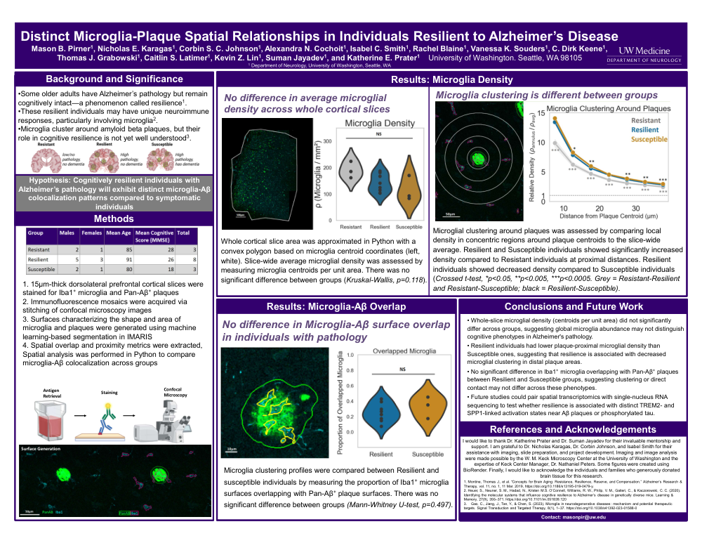
No difference in average microglial density across whole cortical slices
Whole cortical slice area was approximated in Python with a convex polygon based on microglia centroid coordinates (left, white). Slice-wide average microglial density was assessed by measuring microglia centroids per unit area. There was no significant difference between groups (Kruskal-Wallis, p=0.118).
Microglia clustering is different between groups
Microglial clustering around plaques was assessed by comparing local density in concentric regions around plaque centroids to the slice-wide average. Resilient and Susceptible individuals showed significantly increased density compared to Resistant individuals at proximal distances. Resilient individuals showed decreased density compared to Susceptible individuals (Crossed t-test, *p<0.05, **p<0.005, ***p<0.0005. Grey = Resistant-Resilient and Resistant-Susceptible; black = Resilient-Susceptible).
Results: Microglia-Aβ Overlap
No difference in Microglia-Aβ surface overlap in individuals with pathology
Microglia clustering profiles were compared between Resilient and susceptible individuals by measuring the proportion of Iba1⁺ microglia surfaces overlapping with Pan-Aβ⁺ plaque surfaces. There was no significant difference between groups (Mann-Whitney U-test, p=0.497).
Discussion / Conclusions and Future Work
• Whole-slice microglial density (centroids per unit area) did not significantly differ across groups, suggesting global microglia abundance may not distinguish cognitive phenotypes in Alzheimer's pathology.
• Resilient individuals had lower plaque-proximal microglial density than Susceptible ones, suggesting that resilience is associated with decreased microglial clustering in distal plaque areas.
• No significant difference in Iba1⁺ microglia overlapping with Pan-Aβ⁺plaques between Resilient and Susceptible groups, suggesting clustering or direct contact may not differ across these phenotypes.
• Future studies could pair spatial transcriptomics with single-nucleus RNA sequencing to test whether resilience is associated with distinct TREM2- and SPP1-linked activation states near Aβ plaques or phosphorylated tau.
References and Acknowledgements
-
Montine, Thomas J., et al. “Concepts for Brain Aging: Resistance, Resilience, Reserve, and Compensation.” Alzheimer’s Research & Therapy, vol. 11, no. 1, 11 Mar. 2019, https://doi.org/10.1186/s13195-019-0479-y.
-
Heuer, S., Neuner, S. M., Hadad, N., Kristen M.S. O’Connell, Williams, R. W., Philip, V. M., Gaiteri, C., & Kaczorowski, C. C. (2020). Identifying the molecular systems that influence cognitive resilience to Alzheimer’s disease in genetically diverse mice. Learning & Memory, 27(9), 355–371. https://doi.org/10.1101/lm.051839.120
-
Gao, C., Jiang, J., Tan, Y., & Chen, S. (2023). Microglia in neurodegenerative diseases: mechanism and potential therapeutic targets. Signal Transduction and Targeted Therapy, 8(1), 1–37. https://doi.org/10.1038/s41392-023-01588-0
I would like to thank Dr. Katherine Prater and Dr. Suman Jayadev for their invaluable mentorship and support. I am grateful to Dr. Nicholas Karagas, Dr. Corbin Johnson, and Isabel Smith for their assistance with imaging, slide preparation, and project development. Imaging and image analysis were made possible by the W. M. Keck Microscopy Center at the University of Washington and the expertise of Keck Center Manager, Dr. Nathaniel Peters. Some figures were created using BioRender. Finally, I would like to acknowledge the individuals and families who generously donated brain tissue for this research.
Contact
Contact: masonpir@uw.edu
Comments
Email is required but is not shown publicly.
Loading comments...