4,068 posters, 219 videos, 101 topics, 69 sessions, 14,365 authors, 6,128 institutions
ePostersLive by SciGen Technologies S.A. All rights reserved.
Alzheimer's Association International Conference 2026
July 12-15, 2026 | London, United Kingdom, and Online

13430
Vitamin D Modulates Gap-43 And Stmn2 Expression After Axonal Injury
Poster Presenter
Authors
Affiliations
Istanbul University-Cerrahpasa, Brain and Neurodegenerative Disorders Research Laboratories, Department of Neuroscience, Institute of Neurological Sciences, Istanbul, Turkey, Istanbul University-Cerrahpasa, Department of Neuroimmunology, Institute of Neurological Sciences, Istanbul, Turkey
Part of Topic
Basic Science and Pathogenesis -> Molecular and Cell Biology -> Neurodegeneration and neuroprotection
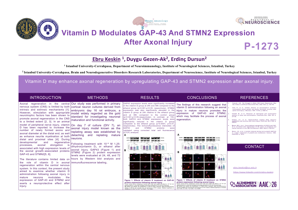
Introduction: Axonal regeneration in the central nervous system (CNS) is limited by both intrinsic and extrinsic mechanisms [1]. However, stimulation with appropriate neurotrophic factors has been shown to promote axonal regeneration in the CNS to a limited extent [2, 3]. In an animal model of peripheral nerve injury, vitamin D has been reported to increase the number of newly formed axons and axonal diameter at the distal end, as well as enhance neurite myelination at both distal and proximal sites [4]. During developmental and regenerative processes, axonal elongation is associated with high expression levels of the axonal growth–associated proteins GAP-43 and STMN2[5, 6]. The literature contains limited data on the role of vitamin D in axonal regeneration within the central nervous system. In this context, the present study aimed to examine whether vitamin D administration following axonal injury in mature neurons modulates the expression of GAP-43 and STMN2 and exerts a neuroprotective effect after injury.
Methods: Our study was performed in primary cortical neuron cultures derived from embryonic day 16 rat embryos, a model widely regarded as the gold standard for investigating neuronal maturation and functional activity. On day 7 of culture (DIV 7), an axonal injury model known as the replating assay was established by detaching and replating mature neurons. Following treatment with 10⁻⁸ M 1,25-dihydroxyvitamin D₃ or ethanol after axonal injury, GAP43 (Figure 1) and STMN2 (Figure 2) protein expression levels were evaluated at 24, 48, and 72 hours by Western blot analysis and immunofluorescence labeling.
Results: GAP43 expression levels were significantly increased in the vitamin D group at 24h and 48h compared to the ethanol group (p<0.001,p<0.05, respectively). STMN2 expression was also significantly elevated in the vitamin D group at 24h relative to the ethanol group and at 48h compared to the control group (p<0.001,p<0.05,respectively). In contrast, at 72h, both GAP43 and STMN2 expression levels were significantly reduced in the vitamin D group compared to the ethanol group (p<0.0001, p<0.01, respectively). The immunofluorescence intensities of GAP43 and STMN2 proteins were validated using corrected total cell fluorescence (CTCF) analysis.
Conclusions: The findings of this research suggest that vitamin D administration following an axonal injury in mature neurons promotes the expression of GAP-43 and STMN2, which may facilitate the process of axonal regeneration.
3. Tetzlaff, W., et al., Response of rubrospinal and corticospinal neurons to injury and neurotrophins. Prog Brain Res, 1994. 103: p. 271-86.
4. Chabas, J.F., et al., Cholecalciferol (vitamin D(3)) improves myelination and recovery after nerve injury. PLoS One, 2013. 8(5): p. e65034.
5. Mason, M.R., et al., Transcriptional upregulation of SCG10 and CAP-23 is correlated with regeneration of the axons of peripheral and central neurons in vivo. Mol Cell Neurosci, 2002. 20(4): p. 595-615.
6. Skene, J.H., et al., A protein induced during nerve growth (GAP-43) is a major component of growth-cone membranes. Science, 1986. 233(4765): p. 783-6.
Comments
Email is required but is not shown publicly.
Loading comments...