4,068 posters, 219 videos, 101 topics, 69 sessions, 14,365 authors, 6,128 institutions
ePostersLive by SciGen Technologies S.A. All rights reserved.
Alzheimer's Association International Conference 2026
July 12-15, 2026 | London, United Kingdom, and Online

13413
Association Of Dysphagia With Thalamic And Cerebellar Volumes In Alzheimer's Disease: A Pilot MRI Study
Poster Presenter
Authors
Affiliations
Part of Topic
Basic Science and Pathogenesis -> Human Neuropathology -> Imaging-pathologic correlations
Association of Dysphagia with Thalamic and Cerebellar Volumes in Alzheimer’s Disease: A Pilot MRI Study
Mihriban Güngör1, Alper Özdemir2, Cemile Özsürekçi3, Ekim Gümeler2, Mustafa Levent1, Elif Gecegelen1, Selen Serel Arslan4, Numan Demir4, Mert Eşme1, Cafer Balcı1, Burcu Balam Doğu1, Mustafa Cankurtaran1, Meltem Gülhan Halil1
1Hacettepe University, Department of Internal Medicine, Division of Geriatric Medicine, Ankara, Turkey
2Hacettepe University, Department of Radiology, Ankara, Turkey
3Lokman Hekim University Hospital, Department of Internal Medicine, Division of Geriatric Medicine, Ankara, Turkey
4Hacettepe University, Faculty of Physical Therapy and Rehabilitation, Ankara, Turkey
Background:
Dysphagia is a critical geriatric syndrome in Alzheimer’s disease (AD) that significantly increases the risk of aspiration pneumonia and mortality. While cortical mechanisms have been investigated, the structural involvement of subcortical regions remains incompletely understood. This exploratory pilot study aimed to investigate the association between dysphagia and subcortical brain volumes in patients with AD.
Methods:
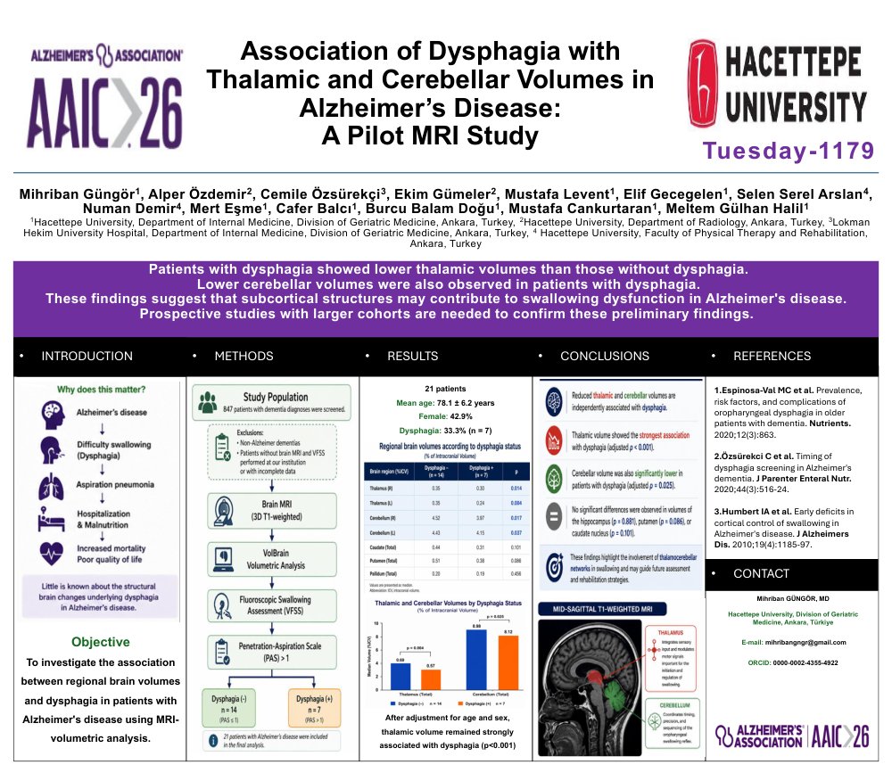
This retrospective cross-sectional pilot study included 21 patients with Alzheimer’s disease who underwent fluoroscopic swallowing assessment between 2016-2018. Dysphagia was defined by clinical symptoms and a Penetration-Aspiration Scale (PAS) score > 1. All patients had brain MRI scans including 3 dimensional T1weighted imaging. Analysis was performed with volbrain software (vol2brain pipeline), volumetric measurements of whole brain structures were obtained. Subcortical brain volumes were compared between patients with and without dysphagia using analysis of covariance (ANCOVA) adjusted for age and sex.
Results:
Among 21 AD patients (mean age 78.14±6.16 years, 42.9% female), 33.3% (n=7) presented with dysphagia. The median interval between MRI and swallowing assessment was 3.3 months (interquartile range: 0.3–13.8 months). There were no significant differences in demographic characteristics between groups. Patients with dysphagia had significantly lower thalamic and cerebellar volumes compared with those without dysphagia (p=0.004 and p=0.025 respectively). After adjustment for age and sex, thalamic volume remained strongly associated with dysphagia (p<0.001), with a large effect size. In contrast, no significant differences were observed in volumes of the hippocampus (p=0.881), putamen (p=0.086), or caudate nucleus (p=0.101) between the groups.
Conclusion:
This exploratory retrospective pilot study demonstrates that dysphagia in AD is associated with reduced thalamic and cerebellar brain volumes. These findings suggest a potential role of thalamic and cerebellar structures in swallowing dysfunction in AD. Larger prospective studies are needed to confirm these preliminary findings.
Keywords: Alzheimer dementia, dysphagia, MRI imaging, PAS score, subcortical brain volumes
References:
- Espinosa-Val MC, Martín-Martínez A, Graupera M, Arias O, Elvira A, Cabré M, Palomera E, Bolívar-Prados M, Clavé P, Ortega O. Prevalence, Risk Factors, and Complications of Oropharyngeal Dysphagia in Older Patients with Dementia. Nutrients. 2020 Mar 24;12(3):863. doi: 10.3390/nu12030863. PMID: 32213845; PMCID: PMC7146553.
Comments
Email is required but is not shown publicly.
Loading comments...