141 posters, 8 videos, 8 audios, 5 topics, 442 authors, 228 institutions
ePostersLive by SciGen Technologies S.A. All rights reserved.
6th World Congress on Vascular Access 2021
April 29 - 30, 2021 | Virtual

P022
Ultrasound-guided hydrodissection for totally implantable central venous access device reservoir pocket: a new procedure for a better outcome
Part of Topic
Education & Training
Ultrasound-guided hydrodissection for totally implantable central venous access device reservoir pocket: a new procedure for a better outcome
F. Longo, F. De Caris, M. Martuscelli, F. Claps, F.E. Agrò
Campus Bio-Medico University Hospital, Anesthesia, Intensive Care and Pain Management Unit.
Rome, Italy
INTRODUCTION
Talking about total implantable central venous access devices (TICVADs), correct positioning of the reservoir is imperative.1 Failing to implant it above the pectoral
muscle, leaving it in the subcutaneous tissue, increases the risk of turning upside down; in addition, the scarce distance from the skin layers increases the risk of
dermal and epidermal erosion.2 Moreover, subcutaneous placement lets the reservoir and the catheter free to move; therefore, risk of damaging the tunica
intima enhances along with the risk of venous thrombosis. Until now, the reservoir pockets have been performed surgically, which does not provide a correct
positioning for certain. We thought of creating the pocket by performing an echoguided hydrodissection with an anesthetic solution, which should guarantee the
right positioning as well as an improvement in postoperative pain, as the tissue trauma caused by surgical instruments is avoided.
METHOD
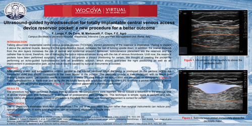
After making a skin and subcutaneous incision parallel to the inferior clavicular edge, the probe is positioned on the pectoral region: the transductor short axis
should correspond to the lower border of the incision. The pectoralis muscle is then visualized, with its fascia and overlying tissue layers. The injection needle is
inserted in in-plane (IP) view through the skin incision and visualized on sonography (Figure 1). Then hydrodissection is madeover and along the pectoralis fascia with a solution of ropivacaine 0.5% to create a pocket immediately above the muscle with no surgical instruments (Figure 2). Port reservoir placement is therefore
performed.
RESULTS
The procedure has been performed in more than 60 patients. No complications were reported. We’ve noticed a reduction in the average time of device implantation and, furthermore, a decrease of postoperative pain complaints. This technique is simple, quick to perform and safe, being totally ultrasound guided.
Timing of procedure is therefore decreased, and reservoir placement is correct for certain.
CONCLUSION
Use of ultrasounds eliminates short-term complications.3 The use of an anesthetic solution rather than surgical instruments can reduce post-operative pain which, in
some kind of patients, can be annoying, especially in fragile ones.