816 posters, 24 topics, 3,314 authors, 1,012 institutions
ePostersLive by SciGen Technologies S.A. All rights reserved.
SAGES 2026 Annual Meeting Emerging Technology Session
March 25-28, 2026 | Tampa, FL, USA

P370
Incidental discovery of diffuse large B cell lymphoma during laparoscopic cholecystectomy:case report
Poster Presenter
Authors
Affiliations
Part of Topic
Biliary
Incidental discovery of diffuse B-cell lymphoma during laparoscopic cholecystectomy: A Case Report
Introduction
Laparoscopic cholecystectomy is one of the most frequently performed minimally invasive surgical procedures and is associated with low morbidity and mortality rates [1]. Postoperative intraperitoneal collections are uncommon and are usually related to bile leakage, bowel injury, or infection. Chylous ascites, defined as the accumulation of triglyceride-rich lymphatic fluid within the peritoneal cavity, is a rare condition most often associated with malignancy, trauma, or lymphatic obstruction [2,3]. Lymphoma is the leading malignant cause of chylous ascites due to lymphatic compression or infiltration [4]. The development of chylous ascites following routine laparoscopic cholecystectomy is exceptional and should prompt investigation for non-surgical etiologies.
Case Presentation
An elderly obese female patient presented with upper abdominal discomfort suggestive of gallstone disease. Her symptoms were atypical and did not follow the classic pattern of biliary colic. She denied fever, jaundice, vomiting, weight loss, or constitutional symptoms. Physical examination was limited by obesity, and no abdominal masses or organomegaly were detected.
Preoperative abdominal ultrasonography demonstrated gallstones with features consistent with chronic calculous cholecystitis, including gallbladder wall thickening and a contracted gallbladder. No intra-abdominal masses or lymphadenopathy were identified. Routine laboratory investigations were within normal limits. Based on these findings, the patient was scheduled for elective laparoscopic cholecystectomy.
Operative Findings
A standard four-port laparoscopic cholecystectomy was performed. The gallbladder appeared chronically inflamed. During exploration, turbid yellowish intraperitoneal fluid was unexpectedly encountered in the subhepatic and pelvic regions. Careful inspection revealed no evidence of bile leak, bowel injury, or visceral perforation. The critical view of safety was achieved, and the cystic duct and artery were secured. The gallbladder was dissected from the liver bed and removed. Given the unexplained intraperitoneal fluid, subhepatic and pelvic drains were placed, and samples were sent for cytology.
Postoperative Course
The immediate postoperative course was initially uneventful. On postoperative day three, following initiation of oral feeding, the drain output increased significantly and became milky white in appearance. Biochemical analysis demonstrated elevated triglyceride levels, confirming chylous ascites [2].
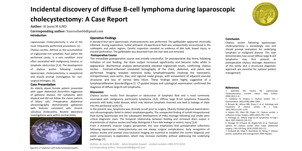
Contrast-enhanced computed tomography of the chest, abdomen, and pelvis was performed. Imaging revealed extensive bulky lymphadenopathy involving the mesenteric, retroperitoneal, para-aortic, iliac, and inguinal nodal groups, with encasement of adjacent vascular structures and loss of normal fatty hilum. These findings were highly suggestive of a lymphoproliferative disorder(figure:1). CT-guided biopsy and cytological examination confirmed the diagnosis of diffuse large B-cell lymphoma.
The patient was referred to the hematology-oncology service for staging and definitive treatment. From a surgical perspective, recovery remained stable, and chylous drainage was managed conservatively with dietary modification and supportive care.
Figure 1: CT abdomen shows bulky lymphadenopathy involving the mesenteric, retroperitoneal para-aortic, iliac, and inguinal nodal groups, with encasement of adjacent vascular structures
Discussion
Chylous ascites results from disruption or obstruction of lymphatic flow and is most commonly associated with malignancy, particularly lymphoma [3,4]. Diffuse large B-cell lymphoma frequently presents with bulky nodal disease, which may obstruct lymphatic channels and lead to leakage of chyle into the peritoneal cavity [5].
In the present case, lymphoma was clinically occult prior to surgery. Obesity limited physical examination, and ultrasonography failed to detect lymphadenopathy. The unexpected finding of turbid intraperitoneal fluid during laparoscopy and the subsequent development of milky drainage following oral intake were critical diagnostic clues. The temporal relationship between feeding and increased drain output is characteristic of chylous ascites and helps distinguish it from bile leakage or enteric injury [2,6].
From a minimally invasive surgery perspective, this case emphasizes that postoperative collections following laparoscopic cholecystectomy are not always surgical complications. Early recognition of chylous ascites and prompt cross-sectional imaging are essential to establish the correct diagnosis and avoid unnecessary re-exploration, which may increase morbidity without addressing the underlying pathology [7].
Conclusion
Chylous ascites following laparoscopic cholecystectomy is exceedingly rare and should prompt evaluation for underlying lymphatic or malignant disease. This case demonstrates how occult diffuse large B-cell lymphoma may first present as postoperative chylous drainage. Awareness of this entity and a structured diagnostic approach are essential for optimal patient management.
Ethics Statement
Written informed consent was obtained from the patient for publication of this case report and accompanying images. Institutional approval was not required for this single case report.
References
1. Sakorafas GH, Tsiotou AG. Laparoscopic cholecystectomy: current status. Surg Endosc. 2000;14(1):1–4.
2. Cárdenas A, Chopra S. Chylous ascites. Am J Gastroenterol. 2002;97(8):1896–1900.
3. Browse NL, Wilson NM, Russo F, et al. Aetiology and treatment of chylous ascites. Br J Surg. 1992;79(11):1145–1150.
4. Aalami OO, Allen DB, Organ CH. Chylous ascites: a collective review. Surgery. 2000;128(5):761–778.
5. Armitage JO. Diffuse large B-cell lymphoma. N Engl J Med. 2007;356(14):139–151.