816 posters, 24 topics, 3,314 authors, 1,012 institutions
ePostersLive by SciGen Technologies S.A. All rights reserved.
SAGES 2026 Annual Meeting Emerging Technology Session
March 25-28, 2026 | Tampa, FL, USA

P366
UTILITY OF INDOCYANINE GREEN (ICG) FLUORESCENCE FOR A PATIENT WITH TRUE LEFT SIDED GALLABLADDER UNDERGOING LAPAROSCOPIC CHOLECYSTECTOMY: A CASE STUDY
Poster Presenter
Authors
Affiliations
Part of Topic
Biliary
Introduction
• True left sided gallbladder (sinistroposition) is rare (0.013 1.1 1 4 Defined as a gallbladder located to the left of the ligamentum teres and falciform ligament
under segment III
• Two variants: associated with situs inversus or left of an aberrantly located right sided round ligament . 5
• Embryologic theories include abnormal hepatic diverticulum attachment or ectopic gallbladder development. 6 7
• Recognition is important as left sided gallbladder increases risk of biliary and vascular anomalies and bile duct injury during laparoscopic cholecystectomy ICG
can be utilized to aid in safer dissection.
Case Presentation
• 56 year old man with HTN, HLD, morbid obesity, insulin dependent T2DM, ESRD on hemodialysis, HFrEF, PVD, CAD s/p CABG, legal blindness, and wheelchair
dependence undergoing evaluation for kidney transplant.
• Presented with 3 months of severe intermittent RUQ pain radiating to right flank. LFTs were normal. Imaging: hepatomegaly, fatty liver, cholelithiasis without
cholecystitis, and gallbladder EF 17% (biliary dyskinesia).
• CT showed left sided gallbladder with a large portal vein branch closely adherent to the posterior gallbladder surface Figure 1.
Operative Details
• Ports modified for left sided gallbladder; not always possible to plan for this preemptively as left sided gallbladder is often an intraoperative finding.
• Gallbladder under segments II III and adherent to falciform ligament Figure 2.
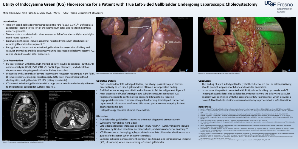
• After dissection of Calot’s triangle, two tubular structures identified; ICG fluorescence used to confirm cystic duct and CBD anatomy Figure 3.
• Large portal vein branch adherent to gallbladder required stapled transection. Laparoscopic ultrasound confirmed biliary and portal venous integrity. Patient
discharged same day
• Histopathology revealed chronic cholecystitis.
Discussion
• True left sided gallbladder is rare and often not diagnosed preoperatively.
• Symptoms may still be right sided.
• Left sided gallbladder increases bile duct injury risk (4.4 7.3%). Variations include abnormal cystic duct insertion, accessory ducts, and aberrant arterial anatomy. 10
• ICG fluorescence cholangiography provides immediate biliary visualization and can guide safe dissection when anatomy is unclear.
• Consider adjusted port placement, surgeon positioning, and intraoperative imaging (ICG, ultrasound) when encountering left sided gallbladder
Conclusion
• The finding of a left sided gallbladder, whether discovered pre or intraoperatively, should prompt suspicion for biliary and vascular anomalies.
• In our case, the patient presented with RUQ pain with biliary dyskinesia and CT imaging showed a left sided gallbladder. Intraoperatively, the biliary and vascular anatomy was confirmed with the assistance of ICG fluorescence, which provides a powerful tool to help elucidate aberrant anatomy to proceed with safe dissection.