816 posters, 24 topics, 3,314 authors, 1,012 institutions
ePostersLive by SciGen Technologies S.A. All rights reserved.
SAGES 2026 Annual Meeting Emerging Technology Session
March 25-28, 2026 | Tampa, FL, USA

P334
Follicular Cholecystitis as a Rare Form of Chronic Cholecystitis: A Lesson in Malignant Mimicry
Part of Topic
Biliary
Follicular Cholecystitis as a Rare Form of Chronic Cholecystitis: A Lesson in Malignant Mimicry
Valerie Fiore, DO1, Jae Hyun Yoo, MS2, Kyle Davis, MS3, Delcasse Joseph, MD1
1Department of General Surgery, Episcopal Health Services; 2Ross University School of Medicine; 3Lake Erie College of Osteopathic Medicine
Introduction:
Follicular cholecystitis (FC) falls under the classification of chronic cholecystitis, and is defined by a histological variant.1 FC has been approximated to be seen in 0.08–11% of cases.2 It has been confused for malignant pathologies such as MALT lymphoma and gallbladder carcinoma, which makes histological verification of utmost importance. Pathologists define FC by three or more lymphoid follicles per centimeter of gallbladder tissue. Not only does this verification aid in guiding treatment plans but may be associated with conditions like primary sclerosing cholangitis, chronic gastritis and pancreatitis.2 We report the case of a 76-year-old woman with chronic gastritis and cholecystitis, found to have FC on pathology following a laparoscopic cholecystectomy, highlighting the need for informed differential diagnosis and acknowledgement of associated conditions.
Case Presentation:
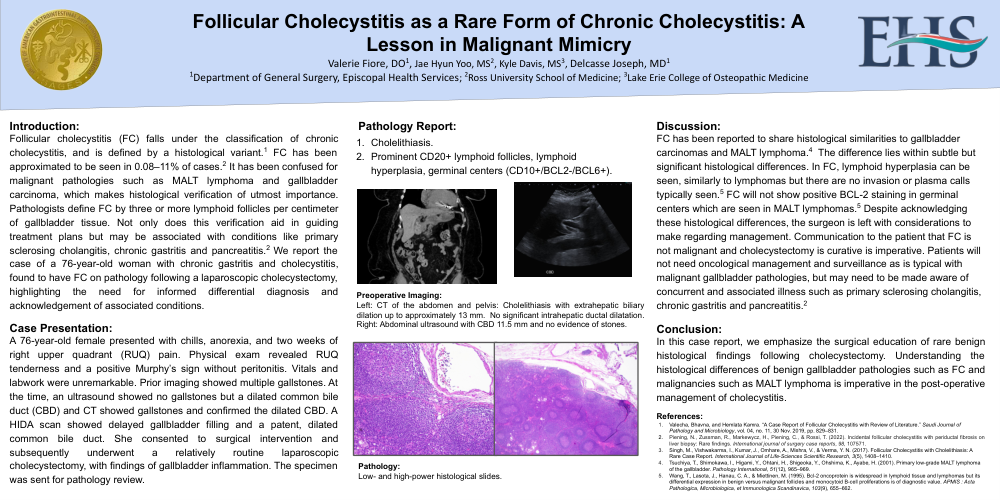
A 76-year-old female presented with chills, anorexia, and two weeks of right upper quadrant (RUQ) pain. Physical exam revealed RUQ tenderness and a positive Murphy’s sign without peritonitis. Vitals and labwork were unremarkable. Prior imaging showed multiple gallstones. At the time, an ultrasound showed no gallstones but a dilated common bile duct (CBD) and CT showed gallstones and confirmed the dilated CBD. A HIDA scan showed delayed gallbladder filling and a patent, dilated common bile duct. She consented to surgical intervention and subsequently underwent a relatively routine laparoscopic cholecystectomy, with findings of gallbladder inflammation. The specimen was sent for pathology review.
Pathology Report:
-
Cholelithiasis.
-
Prominent CD20+ lymphoid follicles, lymphoid hyperplasia, germinal centers (CD10+/BCL2-/BCL6+)
Discussion:
FC has been reported to share histological similarities to gallbladder carcinomas and MALT lymphoma.3 The difference lies within subtle but significant histological differences. In FC, lymphoid hyperplasia can be seen, similarly to lymphomas but there are no invasion or plasma calls typically seen.4 FC will not show positive BCL-2 staining in germinal centers which are seen in MALT lymphomas.4 Despite acknowledging these histological differences, the surgeon is left with considerations to make regarding management. Communication to the patient that FC is not malignant and cholecystectomy is curative is imperative. Patients will not need oncological management and surveillance as is typical with malignant gallbladder pathologies, but may need to be made aware of concurrent and associated illness such as primary sclerosing cholangitis, chronic gastritis and pancreatitis.2
Conclusion:
In this case report, we emphasize the surgical education of rare benign histological findings following cholecystectomy. Understanding the histological differences of benign gallbladder pathologies such as FC and malignancies such as MALT lymphoma is imperative in the post-operative management of cholecystitis.
References:
Valecha, Bhavna, and Hemlata Kamra. "A Case Report of Follicular Cholecystitis with Review of Literature." Saudi Journal of Pathology and Microbiology, vol. 04, no. 11, 30 Nov. 2019, pp. 829–831.
Piening, N., Zussman, R., Markewycz, H., Piening, C., & Rossi, T. (2022). Incidental follicular cholecystitis with periductal fibrosis on liver biopsy: Rare findings. International journal of surgery case reports, 98, 107571.
Tsuchiya, T., Shimokawa, I., Higami, Y., Ohtani, H., Shigeoka, Y., Ohshima, K., Ayabe, H. (2001). Primary low-grade MALT lymphoma of the gallbladder. Pathology International, 51(12), 965–969.
Wang, T., Lasota, J., Hanau, C. A., & Miettinen, M. (1995). Bcl-2 oncoprotein is widespread in lymphoid tissue and lymphomas but its differential expression in benign versus malignant follicles and monocytoid B-cell proliferations is of diagnostic value. APMIS : Acta Pathologica, Microbiologica, et Immunologica Scandinavica, 103(9), 655–662.