2,281 posters, 63 videos, 56 audios, 43 topics, 18 sessions, 6,174 authors, 2,323 institutions
ePostersLive by SciGen Technologies S.A. All rights reserved.
RCOG World Congress 2025
June 23 - 25, 2025 | ExCeL London, UK
EP.0993
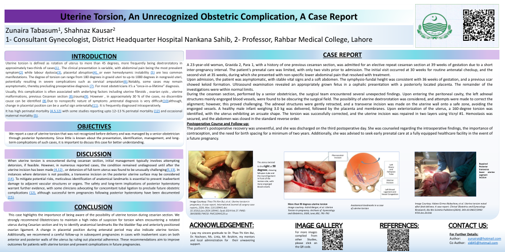
Uterine Torsion:a rare ,unrecognized Obstetric complication.
Poster Presenter
Authors
Affiliations
Part of Topic
High risk pregnancy
Video
INTRODUCTION:
Uterine torsion is defined as rotation of uterus to more than 45 degrees, more frequently being dextrorotatory in approximately two-thirds of cases(1) . The clinical presentation is variable, with abdominal pain being the most prevalent symptom(2) while labour dystocia(3), placental abruption(4), or even hemodynamic instability (5) are less common manifestations. The degree of torsion can range from 180 degrees in gravid uteri to up-to 1080 degrees in nongravid uteri, potentially resulting in severe complications such as cervical amputation(6).Notably, some cases may remain asymptomatic, thereby precluding preoperative diagnosis (7). For most obstetricians it’s a “once-in-a-lifetime” diagnosis.
Usually, this complication is often associated with underlying factors including uterine fibroids , ovarian cysts , uterine malformations, previous Cesarean section (8),trauma(9). However , in approximately 30 % of the cases, no discernible cause can be identified (8).Due to nonspecific nature of symptoms ,antenatal diagnosis is very difficult(10)although, change in placental position can be a useful sign antenatally(11). It is frequently diagnosed intraoperatively.
It has high perinatal mortality (4,5,12) with some studies reporting upto 12–13 % perinatal mortality (12) and occasional maternal mortality (5).
When uterine torsion is encountered during cesarean section, initial management typically involves attempting detorsion, if feasible. However, in numerous reported cases, the condition remained undiagnosed until after the uterine incision has been made (4,12) , or detorsion of full-term uterus was found to be unusually challenging(5,13). In instances where detorsion is not possible, a transverse incision on the posterior uterine surface may be considered (14). To mitigate potential risks, meticulous identification of anatomical landmarks is essential to prevent inadvertent damage to adjacent vascular structures or organs. The safety and long-term implications of posterior hysterotomy warrant further evidence, with some clinicians advocating for concomitant tubal ligation to preclude future obstetric complications (12), although successful term pregnancies following posterior hysterotomy have been documented (15).
CASE REPORT:
A 23-year-old woman, Gravida 2, Para 1, with a history of one previous cesarean section, was admitted for an elective repeat cesarean section at 39 weeks of gestation due to a short inter-pregnancy interval. The patient's prenatal care was limited, with only two visits prior to admission. The initial visit occurred at 30 weeks for routine antenatal checkup, and the second visit at 35 weeks, during which she presented with non-specific lower abdominal pain that resolved with treatment.
Upon admission, the patient was asymptomatic, with stable vital signs and a soft abdomen. The symphysio-fundal height was consistent with 36 weeks of gestation, and a previous scar showed keloid formation. Ultrasound examination revealed an appropriately grown fetus in a cephalic presentation with a posteriorly located placenta. The remainder of the investigations were within normal limits.
During the cesarean section, performed by a senior obstetrician, the surgical team encountered several unexpected findings. Upon entering the peritoneal cavity, the left adnexal structures mainly engorged blood vessels, were found to be obscuring the surgical field. The possibility of uterine dextrorotation was considered, and attempts were made to correct the alignment; however, this proved challenging. The adnexal structures were gently retracted, and a transverse incision was made on the uterine wall onto a safe zone, avoiding the engorged vessels. A healthy male infant weighing 3.0 kg was delivered, followed by the placenta and membranes. Upon exteriorization of the uterus, a 160-degree torsion was identified, with the uterus exhibiting an arcuate shape. The torsion was successfully corrected, and the uterine incision was repaired in two layers using Vicryl #1. Hemostasis was secured, and the abdomen was closed in the standard reverse order.
Postoperative Course and Follow-up:
The patient's postoperative recovery was uneventful, and she was discharged on the third postoperative day. She was counseled regarding the intraoperative findings, the importance of contraception, and the need for birth spacing for a minimum of two years. Additionally, she was advised to seek early prenatal care at a fully equipped healthcare facility in the event of a future pregnancy.
CONCLUSION:
This case highlights the importance of being aware of the possibility of uterine torsion during cesarian section. We strongly recommend Obstetricians to maintain a high index of suspicion for torsion when encountering a rotated uterus during Cesarian section and try to identify anatomical landmarks like the bladder flap and anteriorly positioned ovarian ligament. A change in placental position during antenatal period may also indicate uterine torsion. Additionally, we recommend a careful follow-up in subsequent pregnancies in cases with inadvertent scars on both anterior and posterior walls of the uterus by ruling out placental adherence. These recommendations aim to improve outcomes for patients with uterine torsion and prevent complications in future pregnancies.