2,281 posters, 63 videos, 56 audios, 43 topics, 18 sessions, 6,174 authors, 2,323 institutions
ePostersLive by SciGen Technologies S.A. All rights reserved.
RCOG World Congress 2025
June 23 - 25, 2025 | ExCeL London, UK
EP.0708
Laparoscopic management of ovarian torsion in late pregnancy – Vedio case report
Poster Presenter
Authors
Affiliations
Part of Topic
Gynaecological endoscopy
Video
Laparoscopic management of ovarian torsion in late pregnancy – Video case report
Dr Leena Nagpal (1), Dr Preth De silva (1), Professor T Justin Clark (3), Miss Lina Antoun (2) 1. Registrar in Obstetrics and Gynaecology, 2. Consultant in Obstetrics and Gynaecology, 3. Professor and Consultant in Gynaecology, Birmingham Women’s Hospital, Birmingham, UK.
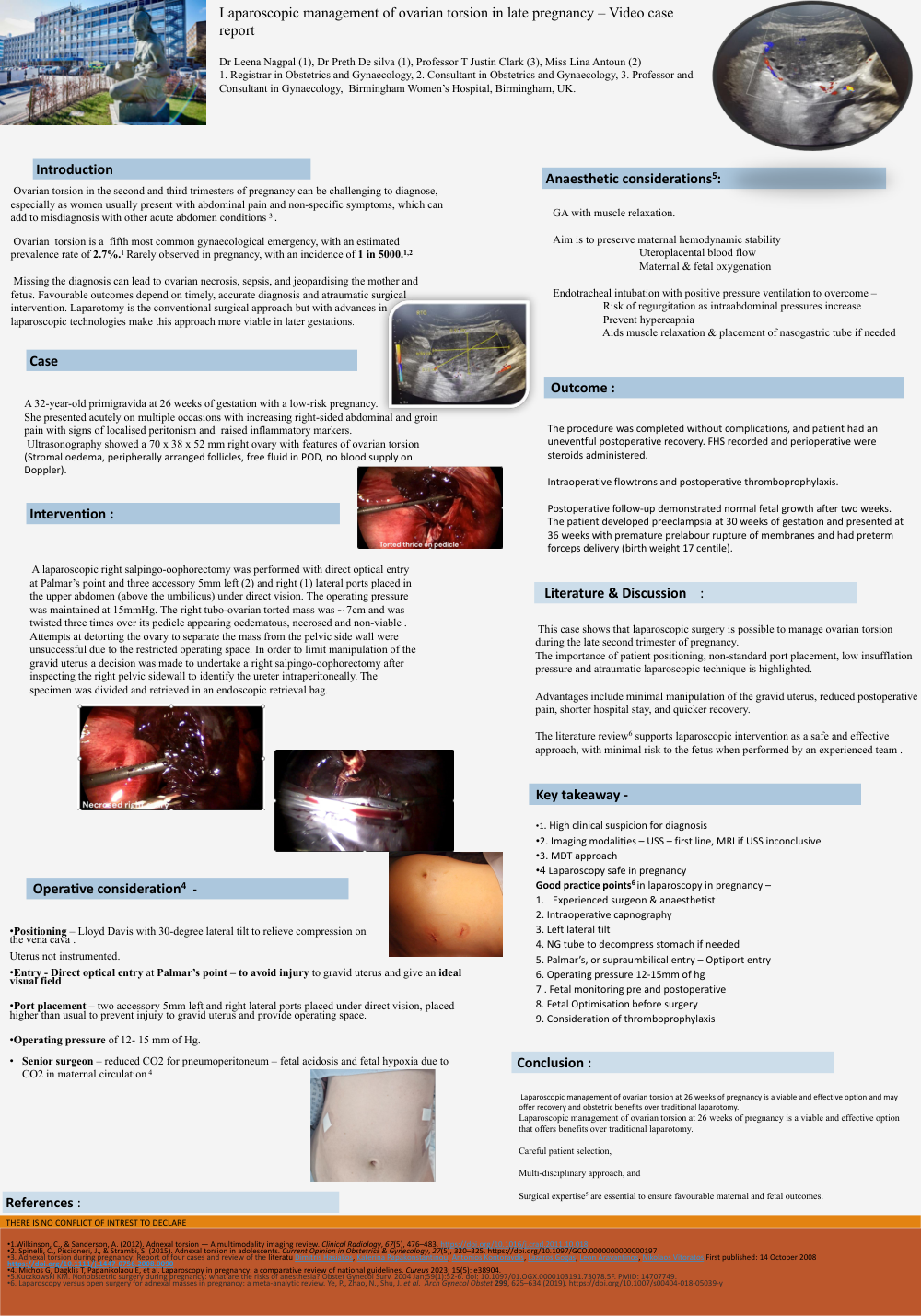
Introduction :Ovarian torsion in the second and third trimesters of pregnancy can be challenging to diagnose, especially as women usually present with abdominal pain and non-specific symptoms, which can add to misdiagnosis with other acute abdomen conditions (3) . Ovarian torsion is a fifth most common gynaecological emergency, with an estimated prevalence rate of 2.7%(1) Rarely observed in pregnancy, with an incidence of 1 in 5000 (1,2) Missing the diagnosis can lead to ovarian necrosis, sepsis, and jeopardising the mother and fetus. Favourable outcomes depend on timely, accurate diagnosis and atraumatic surgical intervention. Laparotomy is the conventional surgical approach but with advances in laparoscopic technologies make this approach more viable in later gestations.
Case Report : A 32-year-old primigravida at 26 weeks of gestation with a low-risk pregnancy. She presented acutely on multiple occasions with increasing right-sided abdominal and groin pain with signs of localised peritonism and raised inflammatory markers. Ultrasonography showed a 70 x 38 x 52 mm right ovary with features of ovarian torsion (Stromal oedema, peripherally arranged follicles, free fluid in POD, no blood supply on Doppler).
Intervention : A laparoscopic right salpingo-oophorectomy was performed with direct optical entry at Palmar’s point and three accessory 5mm left (2) and right (1) lateral ports placed in the upper abdomen (above the umbilicus) under direct vision. The operating pressure was maintained at 15mmHg. The right tubo-ovarian torted mass was ~ 7cm and was twisted three times over its pedicle appearing oedematous, necrosed and non-viable . Attempts at detorting the ovary to separate the mass from the pelvic side wall were unsuccessful due to the restricted operating space. In order to limit manipulation of the gravid uterus a decision was made to undertake a right salpingo-oophorectomy after inspecting the right pelvic sidewall to identify the ureter intraperitoneally. The specimen was divided and retrieved in an endoscopic retrieval bag.
Operative consideration
•Positioning– Lloyd Davis with 30-degree lateral tilt to relieve compression on the vena cava . Uterus not instrumented. •Entry - Direct optical entry at Palmar’s point – to avoid injury to gravid uterus and give an ideal visual field
•Port placement – two accessory 5mm left and right lateral ports placed under direct vision, placed higher than usual to prevent injury to gravid uterus and provide operating space.
•Operating pressure of 12- 15 mm of Hg.
• Senior surgeon – reduced CO2 for pneumoperitoneum – fetal acidosis and fetal hypoxia due to CO2 in maternal circulation (4)
Anaesthetic considerations(5): GA with muscle relaxation. Aim is to preserve maternal hemodynamic stability Uteroplacental blood flow Maternal & fetal oxygenation Endotracheal intubation with positive pressure ventilation to overcome Risk of regurgitation as intraabdominal pressures increase Prevent hypercapnia Aids muscle relaxation & placement of nasogastric tube if needed
Outcome : The procedure was completed without complications, and patient had an uneventful postoperative recovery. FHS recorded and perioperative were steroids administered. Intraoperative flowtrons and postoperative thromboprophylaxis. Postoperative follow-up demonstrated normal fetal growth after two weeks. The patient developed preeclampsia at 30 weeks of gestation and presented at 36 weeks with premature prelabour rupture of membranes and had preterm forceps delivery (birth weight 17 centile).
Literature & Discussion : This case shows that laparoscopic surgery is possible to manage ovarian torsion during the late second trimester of pregnancy. The importance of patient positioning, non-standard port placement, low insufflation pressure and atraumatic laparoscopic technique is highlighted. Advantages include minimal manipulation of the gravid uterus, reduced postoperative pain, shorter hospital stay, and quicker recovery. The literature review6 supports laparoscopic intervention as a safe and effective approach, with minimal risk to the fetus when performed by an experienced team .
Key takeaway •1. High clinical suspicion for diagnosis
•2. Imaging modalities – USS – first line, MRI if USS inconclusive
•3. MDT approach
•4 Laparoscopy safe in pregnancy
Good practice points(6) in laparoscopy in pregnancy
1. Experienced surgeon & anaesthetist
2. Intraoperative capnography
3. Left lateral tilt
4. NG tube to decompress stomach if needed
5. Palmar’s, or supraumbilical entry – Optiport entry
6. Operating pressure 12-15mm of hg
7. Fetal monitoring pre and postoperative
8. Fetal Optimisation before surgery
9. Consideration of thromboprophylaxis
Conclusion :
Laparoscopic management of ovarian torsion at 26 weeks of pregnancy is a viable and effective option and may offer recovery and obstetric benefits over traditional laparotomy. Laparoscopic management of ovarian torsion at 26 weeks of pregnancy is a viable and effective option that offers benefits over traditional laparotomy. Careful patient selection, Multi-disciplinary approach, and Surgical expertise5 are essential to ensure favourable maternal and fetal outcomes.
References : THERE IS NO CONFLICT OF INTREST TO DECLARE
1.Wilkinson, C., & Sanderson, A. (2012). Adnexal torsion — A multimodality imaging review. Clinical Radiology, 67(5), 476–483. https://doi.org/10.1016/j.crad.2011.10.018
2. Spinelli, C., Piscioneri, J., & Strambi, S. (2015). Adnexal torsion in adolescents. Current Opinion in Obstetrics & Gynecology, 27(5), 320–325. https://doi.org/10.1097/GCO.0000000000000197
3. Adnexal torsion during pregnancy: Report of four cases and review of the literatu Dimitris Hasiakos, Katerina Papakonstantinou , Antonios Kontoravdis, Lazaros Gogas , Leon https://doi.org/10.1111/j.1447-0756.2008.0090
4. Michos G, Dagklis T, Papanikolaou E, et al. Laparoscopy in pregnancy: a comparative review of national guidelines. Cureus 2023; 15(5): e38904.
5.Kuczkowski KM. Nonobstetric surgery during pregnancy: what are the risks of anesthesia? Obstet Gynecol Surv. 2004 Jan;59(1):52-6. doi: 10.1097/01.OGX.0000103191.73078.5F. PMID: 14707749. Aravantinos, Nikolaos Vitoratos First published: 14 October 2008
6. Laparoscopy versus open surgery for adnexal masses in pregnancy: a meta-analytic review. Ye, P., Zhao, N., Shu, J. et al. Arch Gynecol Obstet 299, 625–634 (2019). https://doi.org/10.1007/s00404-018-05039-y