2,281 posters, 63 videos, 56 audios, 43 topics, 18 sessions, 6,174 authors, 2,323 institutions
ePostersLive by SciGen Technologies S.A. All rights reserved.
RCOG World Congress 2025
June 23 - 25, 2025 | ExCeL London, UK
EP.0035
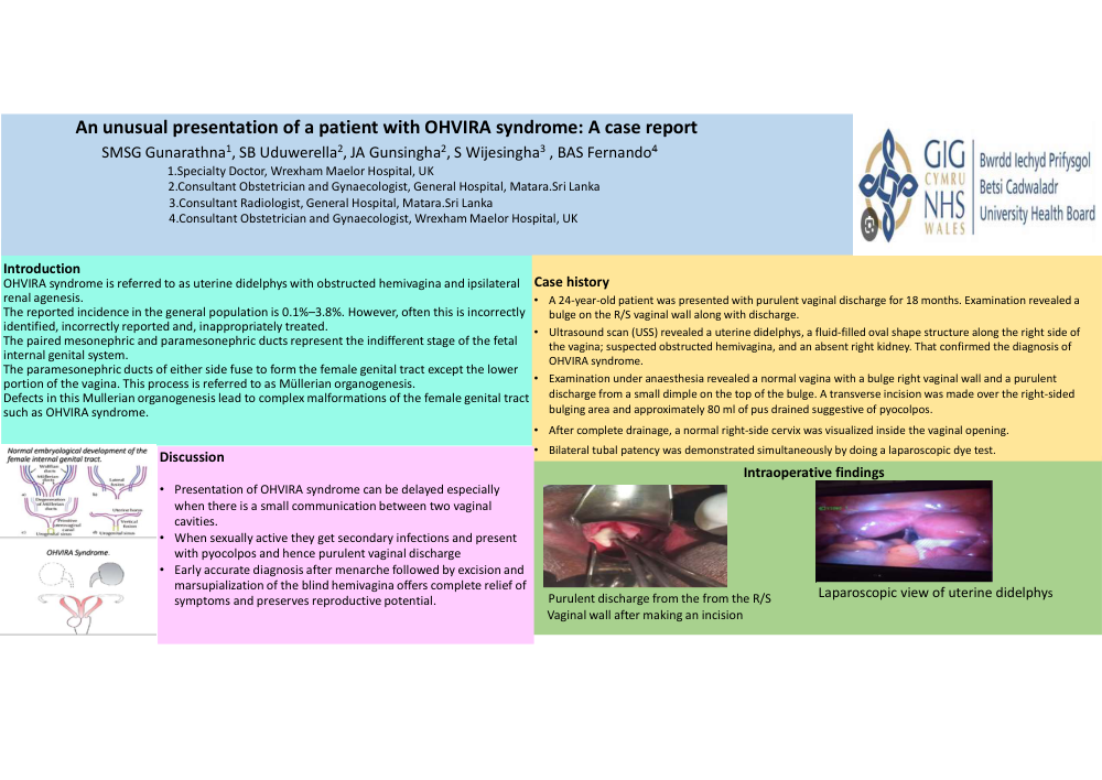
An unusual presentation of a patient with OHVIRA syndrome: A case report
Poster Presenter
Authors
Affiliations
Part of Topic
Adolescent gynaecology
An unusual presentation of a patient with OHVIRA syndrome: A case report
SMSG Gunarathna1, SB Uduwerella2, JA Gunsingha2, S Wijesingha3 , BAS Fernando4
1.Specialty Doctor, Wrexham Maelor Hospital, UK
2.Consultant Obstetrician and Gynaecologist, General Hospital, Matara.Sri Lanka
3.Consultant Radiologist, General Hospital, Matara.Sri Lanka
4.Consultant Obstetrician and Gynaecologist, Wrexham Maelor Hospital, UK
Introduction
OHVIRA syndrome is referred to as uterine didelphys with obstructed hemivagina and ipsilateral renal agenesis.
The reported incidence in the general population is 0.1%–3.8%. However, often this is incorrectly identified, incorrectly reported and, inappropriately treated.
The paired mesonephric and paramesonephric ducts represent the indifferent stage of the fetal internal genital system.
The paramesonephricductsofeithersidefusetoformthefemalegenitaltractexceptthe lower portion of the vagina. This process is referred to as Müllerian organogenesis.
Defects in this Mullerian organogenesis lead to complex malformations of the female genital tract such as OHVIRA syndrome.
Case history
Discussion
Presentation of OHVIRA syndrome can be delayed especially when there is a small communication between two vaginal cavities.
When sexually active they get secondary infections and present with pyocolpos and hence purulent vaginal discharge
Early accurate diagnosis after menarche followed by excision and marsupialization of the blind hemivagina offers complete relief of symptoms and preserves reproductive potential.