2,281 posters, 63 videos, 56 audios, 43 topics, 18 sessions, 6,174 authors, 2,323 institutions
ePostersLive by SciGen Technologies S.A. All rights reserved.
RCOG World Congress 2025
June 23 - 25, 2025 | ExCeL London, UK
EP.0025
Distal Vaginal Atresia: An Atypical Cause of Outflow Tract Obstruction
Poster Presenter
Authors
Affiliations
Part of Topic
Adolescent gynaecology
Distal Vaginal Atresia: An Atypical Cause of Outflow Tract Obstruction
Marc Vision Granada Capunong, RMT, MD; Olive Joyce V. Alabado-Lindero, MD, FPOGS
Adventist Medical Center - Bacolod Department of Obstetrics and Gynecology, Bacolod City, Negros Occidental, Philippines
CASE:
A 17-year-old girl with cyclic hypogastric pain was thought on imaging to have hematometrocolpos with left hematosalpinx from a transverse lower vaginal septum. Intra-op needle mapping yielded no hematocolpos and revealed a tight fibrous plane between bladder and rectum, prompting re-diagnosis as distal vaginal atresia. An ultrasound-guided pull-through vaginoplasty was performed.
OBJECTIVE:
To evaluate the effectiveness and safety of ultrasound-guided pull-through vaginoplasty as a treatment modality for distal vaginal atresia, assessing both short-term outcomes and potential complications.
METHOD:
"ULTRASOUND-GUIDED PULL-THROUGH VAGINOPLASTY"
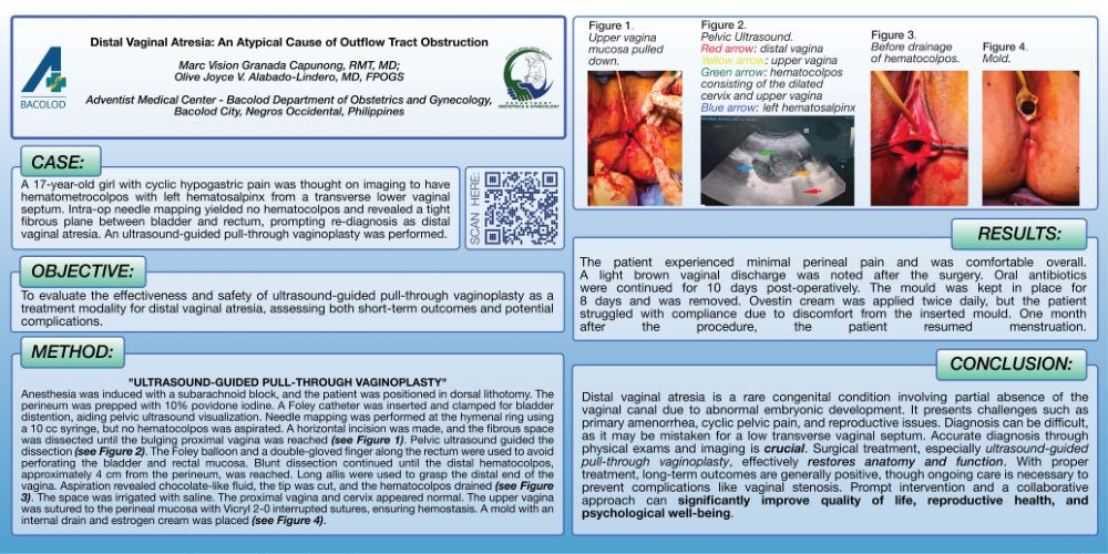
Anesthesia was induced with a subarachnoid block, and the patient was positioned in dorsal lithotomy. The perineum was prepped with 10% povidone iodine. A Foley catheter was inserted and clamped for bladder distention, aiding pelvic ultrasound visualization. Needle mapping was performed at the hymenal ring using a 10 cc syringe, but no hematocolpos was aspirated. A horizontal incision was made, and the fibrous space was dissected until the bulging proximal vagina was reached (see Figure 1). Pelvic ultrasound guided the dissection (see Figure 2). The Foley balloon and a double-gloved finger along the rectum were used to avoid perforating the bladder and rectal mucosa. Blunt dissection continued until the distal hematocolpos, approximately 4 cm from the perineum, was reached. Long allis were used to grasp the distal end of the vagina. Aspiration revealed chocolate-like fluid, the tip was cut, and the hematocolpos drained (see Figure 3). The space was irrigated with saline. The proximal vagina and cervix appeared normal. The upper vagina was sutured to the perineal mucosa with Vicryl 2-0 interrupted sutures, ensuring hemostasis. A mold with an internal drain and estrogen cream was placed (see Figure 4).
RESULTS:
The patient experienced minimal perineal pain and was comfortable overall. A light brown vaginal discharge was noted after the surgery. Oral antibiotics were continued for 10 days post-operatively. The mould was kept in place for 8 days and was removed. Ovestin cream was applied twice daily, but the patient struggled with compliance due to discomfort from the inserted mould. One month after the procedure, the patient resumed menstruation.
CONCLUSION:
Distal vaginal atresia is a rare congenital condition involving partial absence of the vaginal canal due to abnormal embryonic development. It presents challenges such as primary amenorrhea, cyclic pelvic pain, and reproductive issues. Diagnosis can be difficult, as it may be mistaken for a low transverse vaginal septum. Accurate diagnosis through physical exams and imaging is crucial. Surgical treatment, especially ultrasound-guided pull-through vaginoplasty, effectively restores anatomy and function. With proper treatment, long-term outcomes are generally positive, though ongoing care is necessary to prevent complications like vaginal stenosis. Prompt intervention and a collaborative approach can significantly improve quality of life, reproductive health, and psychological well-being.