2,281 posters, 63 videos, 56 audios, 43 topics, 18 sessions, 6,174 authors, 2,323 institutions
ePostersLive by SciGen Technologies S.A. All rights reserved.
RCOG World Congress 2025
June 23 - 25, 2025 | ExCeL London, UK
EP.0032
Vulval Mass in Young Adult – Diagnostic challenge with rare congenital anomaly.
Poster Presenter
Authors
Affiliations
Part of Topic
Adolescent gynaecology
EP.0032
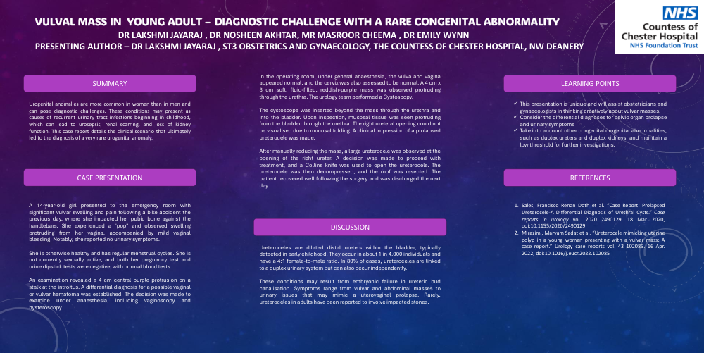
Vulval mass in young adult – Diagnostic Challenge with a rare congenital abnormality
Dr Lakshmi Jayaraj , Dr Nosheen Akhtar, Mr Masroor Cheema , Dr Emily Wynn
Presenting Author – Dr Lakshmi Jayaraj , ST3 Obstetrics and Gynaecology, The Countess of Chester Hospital, NW Deanery
Summary
Urogenital anomalies are more common in women than in men and can pose diagnostic challenges. These conditions may present as causes of recurrent urinary tract infections beginning in childhood, which can lead to urosepsis, renal scarring, and loss of kidney function. This case report details the clinical scenario that ultimately led to the diagnosis of a very rare urogenital anomaly.
Case Presentation
A 14-year-old girl presented to the emergency room with significant vulvar swelling and pain following a bike accident the previous day, where she impacted her pubic bone against the handlebars. She experienced a "pop" and observed swelling protruding from her vagina, accompanied by mild vaginal bleeding. Notably, she reported no urinary symptoms.
She is otherwise healthy and has regular menstrual cycles. She is not currently sexually active, and both her pregnancy test and urine dipstick tests were negative, with normal blood tests.
An examination revealed a 4 cm central purple protrusion on a stalk at the introitus. A differential diagnosis for a possible vaginal or vulvar hematoma was established. The decision was made to examine under anaesthesia, including vaginoscopy and hysteroscopy.
In the operating room, under general anaesthesia, the vulva and vagina appeared normal, and the cervix was also assessed to be normal. A 4 cm x 3 cm soft, fluid-filled, reddish-purple mass was observed protruding through the urethra. The urology team performed a Cystoscopy.
The cystoscope was inserted beyond the mass through the urethra and into the bladder. Upon inspection, mucosal tissue was seen protruding from the bladder through the urethra. The right ureteral opening could not be visualised due to mucosal folding. A clinical impression of a prolapsed ureterocele was made.
After manually reducing the mass, a large ureterocele was observed at the opening of the right ureter. A decision was made to proceed with treatment, and a Collins knife was used to open the ureterocele. The ureterocele was then decompressed, and the roof was resected. The patient recovered well following the surgery and was discharged the next day.
Discussion
Ureteroceles are dilated distal ureters within the bladder, typically detected in early childhood. They occur in about 1 in 4,000 individuals and have a 4:1 female-to-male ratio. In 80% of cases, ureteroceles are linked to a duplex urinary system but can also occur independently.
These conditions may result from embryonic failure in ureteric bud canalisation. Symptoms range from vulvar and abdominal masses to urinary issues that may mimic a uterovaginal prolapse. Rarely, ureteroceles in adults have been reported to involve impacted stones.
Learning Points
ü This presentation is unique and will assist obstetricians and gynaecologists in thinking creatively about vulvar masses.
ü Consider the differential diagnoses for pelvic organ prolapse and urinary symptoms
ü Take into account other congenital urogenital abnormalities, such as duplex ureters and duplex kidneys, and maintain a low threshold for further investigations.
References
- Sales, Francisco Renan Doth et al. “Case Report: Prolapsed Ureterocele-A Differential Diagnosis of Urethral Cysts.” Case reports in urology vol. 2020 2490129. 18 Mar. 2020, doi:10.1155/2020/2490129
- Mirazimi, Maryam Sadat et al. “Ureterocele mimicking uterine polyp in a young woman presenting with a vulvar mass: A case report.” Urology case reports vol. 43 102085. 16 Apr. 2022, doi:10.1016/j.eucr.2022.102085