2,281 posters, 63 videos, 56 audios, 43 topics, 18 sessions, 6,174 authors, 2,323 institutions
ePostersLive by SciGen Technologies S.A. All rights reserved.
RCOG World Congress 2025
June 23 - 25, 2025 | ExCeL London, UK
EP.0272
Ovarian Ectopic Pregnancy: Presentation and Management
Poster Presenter
Part of Topic
Early pregnancy complications/early pregnancy and acute gynaecology
The Presentation and Management of Ovarian Ectopic Pregnancy: A Case Series
Dr Areeba Jawaid, Dr Taona Nyamapfene, Dr Amr Mohammed, Ms Kristin Fiedler
Royal Cornwall Hospital Trust
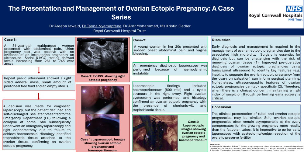
Case 1:
A 31-year-old multiparous woman presented with abdominal pain. Urine pregnancy test was positive with no evidence of an intrauterine pregnancy on ultrasound. Serial β-hCG testing showed levels increasing from 261 to 745 over 48hrs.
Repeat pelvic ultrasound showed a right sided adnexal mass, small amount of peritoneal free fluid and an empty uterus.
A decision was made for diagnostic laparoscopy, but the patient declined and self-discharged. She later presented to the Emergency Department (ED) following a collapse at home. She subsequently underwent an emergency laparoscopy and right oophorectomy due to failure to achieve haemostasis. Histology identified trophoblastic tissue attached to the ovarian tissue, confirming an ovarian ectopic pregnancy.
Image from Case 1: TVUSS showing right ectopic pregnancy
Image from Case 1: Laparoscopic images showing ovarian ectopic pregnancy and haemoperitoneum
Case-2:
A young woman in her 20s presented with sudden onset abdominal pain and vaginal bleeding.
An emergency diagnostic laparoscopy was performed because of haemodynamic instability.
Laparoscopic findings included haemoperitoneum (800 mls) and a cystic structure in the right ovary. Right ovarian cystectomy was performed, and histology confirmed an ovarian ectopic pregnancy with the presence of chorionic-villi and trophoblastic tissue.
Image from Case 2: Laparoscopic images showing ovarian ectopic pregnancy and haemoperitoneum
Discussion
Early diagnosis and management is required in the management of ovarian ectopic pregnancies due to the associated high morbidity. Surgery is essential for diagnosis but can be challenging with the risk of removing ovarian tissue (1). Improved pre-operative diagnosis of ovarian ectopic pregnancies using transvaginal ultrasound to identify key features (e.g. inability to separate the ovarian ectopic pregnancy from the ovary on palpation) can inform surgical planning. Nonetheless, ultrasonographic features of ovarian ectopic pregnancies can lack specificity (2). Therefore, when there is a clinical concern, maintaining a high index of suspicion through performing early surgery is critical.
Conclusion
The clinical presentation of tubal and ovarian ectopic pregnancies may be similar. Still, ovarian ectopic pregnancies often remain asymptomatic as the ovary compensates for the growing pregnancy more readily than the fallopian tubes. It is imperative to go for early laparoscopy with cystectomy/wedge resection of the ovary to preserve fertility.
References
1. Solangon SA, Naftalin J, Jurkovic D. Ovarian ectopic pregnancy: clinical characteristics, ultrasound diagnosis and management. Ultrasound in Obstetrics Gynecology. 2024;63(6):815-23. 2. Sosa M, Brancazio S, Drummey A, Nguyen T, Toussaint T. Transvaginal Ultrasound Diagnosis of Ovarian Ectopic Pregnancy. Cureus. 2023;15(1):e33536.