2,281 posters, 63 videos, 56 audios, 43 topics, 18 sessions, 6,174 authors, 2,323 institutions
ePostersLive by SciGen Technologies S.A. All rights reserved.
RCOG World Congress 2025
June 23 - 25, 2025 | ExCeL London, UK
EP.0115
Splenic Ectopic Pregnancy - A Diagnostic Challenge
Poster Presenter
Authors
Yomna Fahmy, Claire Ross, Nora Kiss, Rasana Bajracharya, Duaa Gumaa, Ee Xuan Ngeyu
Affiliations
Part of Topic
Early pregnancy complications/early pregnancy and acute gynaecology
Splenic Ectopic Pregnancy – A Diagnostic Challenge
Yomna Fahmy – Maidstone and Tunbridge Wells NHS Trust
Introduction
Ectopic pregnancy occurs in 1-2% of pregnancies with 95% being in the fallopian tubes, thus the presence of a splenic ectopic pregnancy is exceedingly rare and poses significant diagnostic and therapeutic challenges. This is a case report of a rare case of ruptured splenic ectopic pregnancy.
Case report
A 36-year-old P3(uneventful pregnancies, all SVD) attended the Emergency Gynae Assessment unit with abdominal pain, no PV bleeding and a positive pregnancy test. PSH: Appendectomy., no PMH. Allergic to Penicillin.
Initial investigations showed: B-HCG 24076, Progesterone 14, Hb 130, CRP 9, with normal LFTs and U&Es .
TVS: showed an empty uterine cavity, a small cystic structure in the right adnexa possibly an ectopic vs fimbrial cyst. With this presentation and no intrauterine pregnancy seen on the TVS and elevated BHCG, an ectopic pregnancy was suspected, and a diagnostic laparoscopy was performed.
Laparoscopy: a right side fimbrial cyst was seen with otherwise normal fallopian tubes on both sides. 300ml of blood was noted in the pelvis but no source of bleeding identified, and no ectopic pregnancy seen in the pelvis. The abdominal structures were examined but no abnormality identified, and a tubal miscarriage was suspected and the fimbrial cyst removed. The plan was for a repeat B-HCG the following day (48 hours from the last one) to assess resolution of pregnancy.
Day 1 post-laparoscopy: The patient was complaining of worsening pain, repeat bloods showed BHCG 23769, Hb 97, CRP 8, normal LFTS and U&Es. Given the significant drop in Hb, inconsistent with the blood loss at laparoscopy and the very high BHCG with no sign of intrauterine pregnancy a decision was made for urgent CT to look for other possible sites of ectopic pregnancy and the source of bleeding
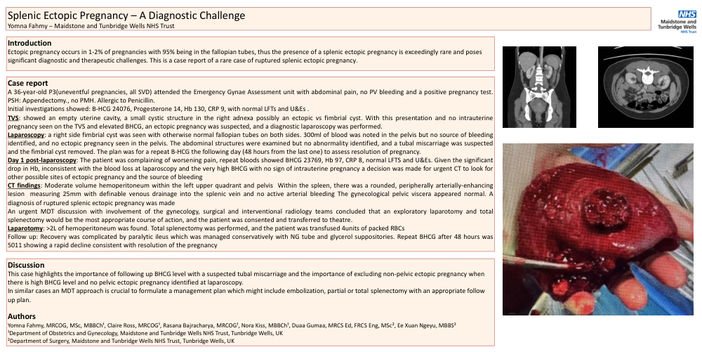
CT findings: Moderate volume hemoperitoneum within the left upper quadrant and pelvis Within the spleen, there was a rounded, peripherally arterially-enhancing lesion measuring 25mm with definable venous drainage into the splenic vein and no active arterial bleeding The gynecological pelvic viscera appeared normal. A diagnosis of ruptured splenic ectopic pregnancy was made
An urgent MDT discussion with involvement of the gynecology, surgical and interventional radiology teams concluded that an exploratory laparotomy and total splenectomy would be the most appropriate course of action, and the patient was consented and transferred to theatre.
Laparotomy: >2L of hemoperitoneum was found. Total splenectomy was performed, and the patient was transfused 4units of packed RBCs
Follow up: Recovery was complicated by paralytic ileus which was managed conservatively with NG tube and glycerol suppositories. Repeat BHCG after 48 hours was 5011 showing a rapid decline consistent with resolution of the pregnancy
Discussion
This case highlights the importance of following up BHCG level with a suspected tubal miscarriage and the importance of excluding non-pelvic ectopic pregnancy when there is high BHCG level and no pelvic ectopic pregnancy identified at laparoscopy.
In similar cases an MDT approach is crucial to formulate a management plan which might include embolization, partial or total splenectomy with an appropriate follow up plan.
Authors
Yomna Fahmy, MRCOG, MSc, MBBCh¹, Claire Ross, MRCOG¹, Rasana Bajracharya, MRCOG¹, Nora Kiss, MBBCh¹, Duaa Gumaa, MRCS Ed, FRCS Eng, MSc², Ee Xuan Ngeyu, MBBS²
¹Department of Obstetrics and Gynecology, Maidstone and Tunbridge Wells NHS Trust, Tunbridge Wells, UK
²Department of Surgery, Maidstone and Tunbridge Wells NHS Trust, Tunbridge Wells, UK