2,281 posters, 63 videos, 56 audios, 43 topics, 18 sessions, 6,174 authors, 2,323 institutions
ePostersLive by SciGen Technologies S.A. All rights reserved.
RCOG World Congress 2025
June 23 - 25, 2025 | ExCeL London, UK
EP.0268
Chorionic Bump: A Potential Diagnostic Pitfall in the First Trimester
Poster Presenter
Authors
Sonal Changal, Pushpee Sinha, Shona Bhome-Dhaliwal, Melissa Islam, Iranthi Meththananda
Affiliations
Part of Topic
Early pregnancy complications/early pregnancy and acute gynaecology
Title: Chorionic Bump: A Potential Diagnostic Pitfall in the First Trimester
Objective: This case report details the incidental ultrasound detection of a chorionic bump in a 30-year-old G2P0 patient at 6+3 weeks of gestation. Chorionic bumps are an increasingly recognized sonographic finding in early pregnancy and have been associated with an elevated risk of early pregnancy loss. However, in ongoing pregnancies, these structures typically resolve spontaneously without any deleterious effects on fetal development.
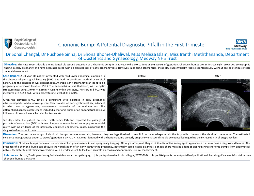
Case Report: A 30-year-old patient presented with mild lower abdominal cramping in the absence of per vaginal bleeding (PVB). She had no significant medical or surgical history, and the conception was spontaneous. An initial early pregnancy scan identified a pregnancy of unknown location (PUL). The endometrium was thickened, with a cystic structure measuring 1.9mm × 3.8mm × 7.8mm within the cavity. Her serum β-hCG was measured at 12,858 IU/L, with a progesterone level of 38 nmol/L.
Given the elevated β-hCG levels, a consultant with expertise in early pregnancy ultrasound performed a follow-up scan. This revealed an early gestational sac, adjacent to which was a hyperechoic, non-vascular protrusion of the endometrium. The differential diagnoses at this stage included a chorionic bump or an endometrial polyp. A follow-up ultrasound was scheduled for two weeks.
Ten days later, the patient presented with heavy PVB and reported the passage of products of conception (POC) at home. A repeat scan confirmed an empty endometrial cavity, with no evidence of the previously visualised endometrial mass, supporting the diagnosis of a chorionic bump.
Discussion: The precise aetiology of chorionic bumps remains uncertain; however, they are hypothesized to result from hemorrhage within the trophoblast beneath the chorionic membrane. The estimated incidence in pregnancies under 10 weeks' gestation is 0.4–0.7%. Patients identified with a chorionic bump on early pregnancy ultrasound should be counselled regarding the increased risk of pregnancy loss.
Conclusions: Chorionic bumps remain an under-researched phenomenon in early pregnancy imaging. Although infrequent, they exhibit a distinctive sonographic appearance that may pose a diagnostic dilemma. The presence of a chorionic bump can obscure the visualization of an early intrauterine pregnancy, potentially complicating diagnosis. Sonographers must be adept at distinguishing chorionic bumps from endometrial polyps, the latter typically being hyperechoic with a feeder vessel, to facilitate accurate diagnosis and appropriate clinical management.