2,281 posters, 63 videos, 56 audios, 43 topics, 18 sessions, 6,174 authors, 2,323 institutions
ePostersLive by SciGen Technologies S.A. All rights reserved.
RCOG World Congress 2025
June 23 - 25, 2025 | ExCeL London, UK
EP.0267
An atypical presentation of ectopic pregnancy in a 42 year old female
Part of Topic
Early pregnancy complications/early pregnancy and acute gynaecology
AN ATYPICAL PRESENTATION OFECTOPICPREGNANCY IN A 42YEAR OLD FEMALE:
INTRODUCTION
Ectopic pregnancy, defined as a pregnancy occurring outside the uterine cavity` occurs in 11/1000 pregnancies in the UK with an estimated 11000 ectopic pregnancies diagnosed each year. Ectopic pregnancy remains a leading cause of maternal death, and a delayed diagnosis is associated with significant morbidity and mortality(1).
DISCUSSION
Ectopic pregnancy is a high-risk condition: a leading cause of maternal mortality in the first trimester with an incidence of 5-10% of all pregnancy related deaths. It may present a diagnostic challenge in the emergency department. While the typical presentation of ectopic pregnancy includes a triad of amenorrhea, abdominal pain, and vaginal bleeding, these symptoms occur in only one-third of cases (2).
CASE PRESENTATION
A 42-year-old woman (G11P5+5) presented to Creek General Hospital with two weeks of cyclical vaginal bleeding and lower abdominal pain, along with one week of dysuria. Her history included irregular contraceptive use (oral, injectable, emergency, and IUDs), five induced miscarriages, and poorly controlled type 2 diabetes. Her last menstrual period was July 3, 2022, with ongoing spotting followed by heavy flow. She denied amenorrhea.
EXAMINATION
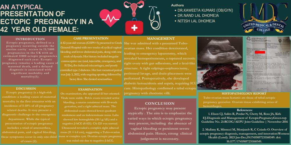
On examination, she appeared ill but oriented. Vitals were stable. Pelvic exam revealed active bleeding, a uterus consistent with 10-week gestation, and a right adnexal mass. The abdominal exam showed lower quadrant tenderness and an indeterminate mass. Labs showed low hemoglobin (10.1 g/dL) and a negative β-hCG (0.421). CA-125 was normal. Ultrasound revealed a complex right adnexal mass (11 × 6.8 cm), suggesting a Tubo-ovarian mass or complex cyst. Ovarian ectopic pregnancy was ruled out due to negative β-hCG.
MANAGEMENT
She was admitted with a presumed Tubo-ovarian mass. Her condition deteriorated, leading to emergency laparotomy, which revealed hemoperitoneum, a ruptured necrotic right ovary with gut adhesions, and a fetal-like structure. A right salpingo-oophorectomy, peritoneal lavage, and drain placement were performed. Postoperatively, she developed diabetic ketoacidosis and sepsis, requiring ICU care. Histopathology confirmed a tubal ectopic pregnancy with chorionic villi.
HISTOPATHOLOGY REPORT
Tubo-ovarian mass features are those of tubal ectopic
pregnancy gestationOvarian tissue exhibiting areas of
hemorrhages.
CONCLUSION
Ectopic pregnancy may present atypically . The aim is to emphasize the varied ways in which ectopic pregnancy may present, includingthe absence of vaginal bleeding or persistent severe abdominal pain. Hence, strongclinical judgement is necessary.
References:
1. Elson CJ, Salim R, Potdar N, Chetty M, Ross JA, Kirk EJ.Diagnosis and Management of EctopicPregnancyGreen-top Guideline No. 21:RCOG/AEPU Joint Guideline | November 2016
2. Mullany K, Minneci M, Monjazeb R, C Coiado O. Overview of ectopic pregnancy diagnosis, management, and innovation:Womens Health (Lond). 2023 Jan-Dec;19:17455057231160349. doi: 10.1177/17455057231160349.
Authors
- DR.KAWEETA KUMARI (OB/GYN)
- DR.NAND LAL DHOMEJA
- NITISH LAL DHOMEJA