2,281 posters, 63 videos, 56 audios, 43 topics, 18 sessions, 6,174 authors, 2,323 institutions
ePostersLive by SciGen Technologies S.A. All rights reserved.
RCOG World Congress 2025
June 23 - 25, 2025 | ExCeL London, UK
EP.0261
Case Reports on IUCD-Related Uterine Perforation: Prevention, Early Recognition, and Management
Poster Presenter
Authors
Faozia Wangde, Zil E Haider Syed, Fiza Akram, Maged Shendy, Hina Ejaz
Affiliations
Part of Topic
Early pregnancy complications/early pregnancy and acute gynaecology
Objectives:
The objective of this report is to emphasize:
• The importance of adhering to safe IUCD insertion
techniques,
• Early recognition and diagnosis of IUCD complications,
• The role of multidisciplinary management
• Evidence-based prevention and follow-up strategies.
These case reports illustrate how prompt imaging, surgical
planning, and interdisciplinary collaboration can prevent
severe outcomes like bowel injury or sepsis.
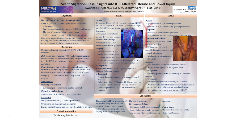
Case 1
Patient
51-year-old, History of endometriosis; previous LLETZ
for CIN 3, Copper IUCD (Cu-IUCD) inserted in hospital
after failed GP attempt (under local anaesthesia)
Symptoms
Severe, acute lower-abdominal
pain 2 weeks post-insertion
Examination Findings
Pelvic tenderness; IUCD
threads not visible on speculum
Imaging
• Pelvic ultrasound: IUCD
• absent from uterine cavity
• CT/MRI: device located in
• pelvis, adherent to uterosacral ligament and in close
apposition to sigmoid colon with dense adhesions
Management
a. Multidisciplinary Team : Gynaecologist (lead
surgeon), Colorectal surgeon
b. Treatment
• Planned diagnostic laparoscopy with pre-op bowel
prep.
• Adhesiolysis done; IUCD embedded between uterine
serosa and sigmoid colon, no bowel perforation
• Laparoscopic retrieval of IUCD; bowel intact - no
resection
Case 2
Patient
26-year-old woman, Six months postpartum
Underwent Cu-IUCD insertion by general practitioner
Symptoms
--Immediate discomfort during insertion
--Later presented with abdominal pain and signs of sepsis
Examination Findings
--Suspected uterine perforation
at time of insertion
--IUCD threads visualized
embedded in anterior uterine wall
during hysteroscopy
--Failed retrieval attempt during
hysteroscopy
Imaging
• MRI confirmed full-thickness uterine perforation
• IUCD located adjacent to the sigmoid colon
Management
a. Multidisciplinary Team: Gynaecologist, Colorectal
surgeon
b. Treatment
• Emergency diagnostic laparoscopy due to sepsis
• IUCD partially embedded in anterior myometrium and in
contact with sigmoid serosa, no bowel perforation.
• Adhesiolysis and laparoscopic retrieval of IUCD
Discussion
Uterine perforation occurs in 0.1–0.3% of IUCD
insertions.
Risk higher in postpartum (<36 weeks), breastfeeding,
uterine anomalies, retroverted uterus, surgical history
(e.g., LLETZ), and insertions by less experienced
providers.
Complications: Can lead to adhesions, chronic pain,
infertility, or organ injury. 15% of perforations involve
bowel or bladder. Bowel involved in 3–7.5% of cases.
Imaging: Ultrasound first-line; X-ray or MRI for migrated
Management
Partial perforations:
--hysteroscopic or ultrasound-guided removal.
Complete perforations:
--laparoscopy with pre-op bowel preparation.
Prevention
Delay insertion until ≥12 weeks postpartum.
Ultrasound guidance in high-risk cases.
Ensure proper training and post-insertion follow-up with
patient education.
Conclusions
These cases highlight importance of
prevention, early detection, and
multidisciplinary management in
IUCD related uterine perforation,
helping avoid major complications
like bowel resection.
Key recommendations:
•Standardize IUCD insertion training
•Ensure follow-up in high-risk cases
•Use imaging when complications are
suspected
Future focus:
•Post-insertion ultrasound utility
•Safer IUCD designs