2,281 posters, 63 videos, 56 audios, 43 topics, 18 sessions, 6,174 authors, 2,323 institutions
ePostersLive by SciGen Technologies S.A. All rights reserved.
RCOG World Congress 2025
June 23 - 25, 2025 | ExCeL London, UK
EP.0251
A rare case of sigmoidal ectopic pregnancy
Poster Presenter
Part of Topic
Early pregnancy complications/early pregnancy and acute gynaecology
A Rare Case of Sigmoidal Ectopic Pregnancy: A Case Report
Objective:
Abdominal ectopic pregnancies (AEPs) are an extremely rare but significant form of ectopic pregnancy, accounting for less than 1% of ectopic pregnancies and with a maternal mortality rate estimated to be 7.7 times higher than that of other locations of ectopic pregnancy. They occur when the fertilized egg implants outside the uterus and within the abdominal cavity. In view of the rarity of the condition and the lack of consensus on best treatment modality, we herein report a complex case of a sigmoid ectopic pregnancy.
Case report:
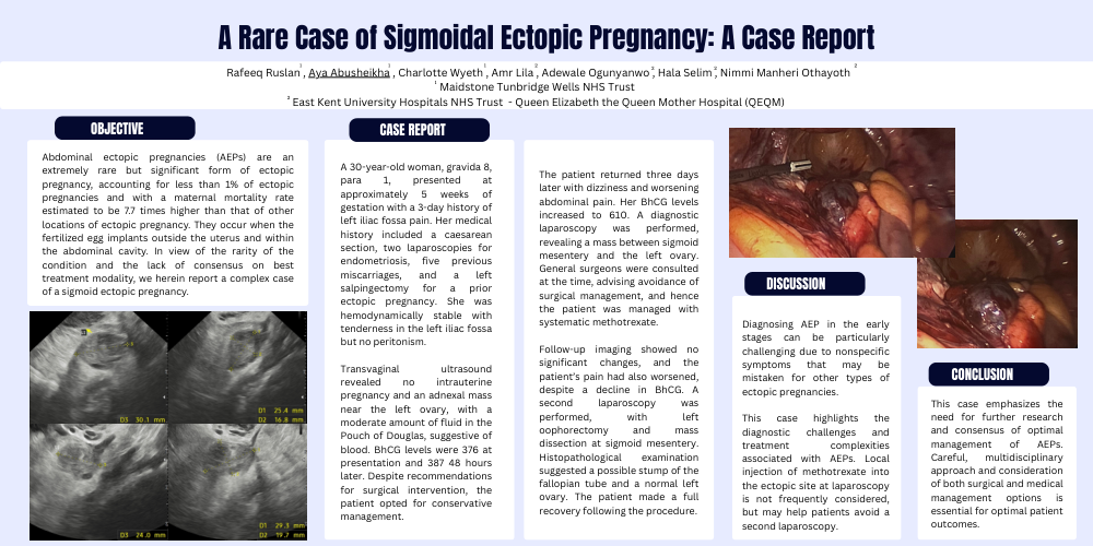
A 30-year-old woman, gravida 8, para 1, presented at approximately 5 weeks of gestation with a 3-day history of left iliac fossa pain. Her medical history included a caesarean section, two laparoscopies for endometriosis, five previous miscarriages, and a left salpingectomy for a prior ectopic pregnancy. She was hemodynamically stable with tenderness in the left iliac fossa but no peritonism. Transvaginal ultrasound revealed no intrauterine pregnancy and an adnexal mass near the left ovary, with a moderate amount of fluid in the Pouch of Douglas, suggestive of blood. BhCG levels were 376 at presentation and 387 48 hours later. Despite recommendations for surgical intervention, the patient opted for conservative management.
The patient returned three days later with dizziness and worsening abdominal pain. Her BhCG levels increased to 610. A diagnostic laparoscopy was performed, revealing a mass between sigmoid mesentery and the left ovary. General surgeons were consulted at the time, advising avoidance of surgical management, and hence the patient was managed with systematic methotrexate. Follow-up imaging showed no significant changes, and the patient’s pain had also worsened, despite a decline in BhCG. A second laparoscopy was performed, with left oophorectomy and mass dissection at sigmoid mesentery. Histopathological examination suggested a possible stump of the fallopian tube and a normal left ovary. The patient made a full recovery following the procedure.
Discussion:
Diagnosing AEP in the early stages can be particularly challenging due to nonspecific symptoms that may be mistaken for other types of ectopic pregnancies. This case highlights the diagnostic challenges and treatment complexities associated with AEPs. Local injection of methotrexate into the ectopic site at laparoscopy is not frequently considered, but may help patients avoid a second laparoscopy.
Conclusion:
This case emphasizes the need for further research and consensus of optimal management of AEPs. Careful, multidisciplinary approach and consideration of both surgical and medical management options is essential for optimal patient outcomes.