2,281 posters, 63 videos, 56 audios, 43 topics, 18 sessions, 6,174 authors, 2,323 institutions
ePostersLive by SciGen Technologies S.A. All rights reserved.
RCOG World Congress 2025
June 23 - 25, 2025 | ExCeL London, UK
EP.0241
Final confirmation of a rare ectopic pregnancy at laparoscopy
Poster Presenter
Part of Topic
Early pregnancy complications/early pregnancy and acute gynaecology
Final confirmation of a rare ectopic pregnancy at laparoscopy
Stephanie Balogun1, Alina-Maria Unipan1
1Queen’s Hospital, Romford
Case Summary
A 31 year old nulliparous women, attended our local early pregnancy unit at 6+3 weeks gestation with a positive pregnancy test, a history of abdominal pain and moderate vaginal bleeding for 3 days.
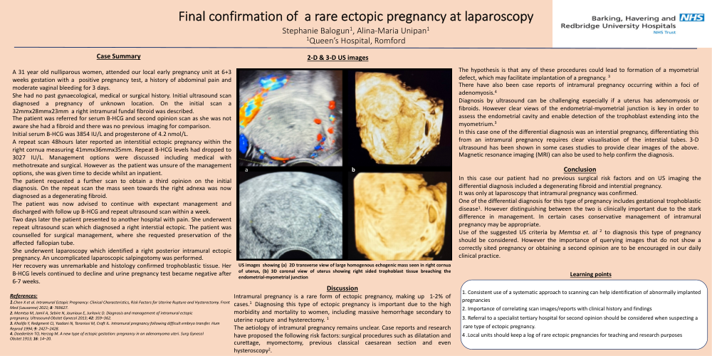
She had no past gynaecological, medical or surgical history. Initial ultrasound scan diagnosed a pregnancy of unknown location. On the initial scan a 32mmx28mmx23mm a right intramural fundal fibroid was described.
The patient was referred for serum B-HCG and second opinion scan as she was not aware she had a fibroid and there was no previous imaging for comparison.
Initial serum B-HCG was 3854 IU/L and progesterone of 4.2 nmol/L.
A repeat scan 48hours later reported an interstitial ectopic pregnancy within the right cornua measuring 41mmx36mmx35mm. Repeat B-HCG levels had dropped to 3027 IU/L. Management options were discussed including medical with methotrexate and surgical. However as the patient was unsure of the management options, she was given time to decide whilst an inpatient.
The patient requested a further scan to obtain a third opinion on the initial diagnosis. On the repeat scan the mass seen towards the right adnexa was now diagnosed as a degenerating fibroid.
The patient was now advised to continue with expectant management and discharged with follow up B-HCG and repeat ultrasound scan within a week.
Two days later the patient presented to another hospital with pain. She underwent repeat ultrasound scan which diagnosed a right interstial ectopic. The patient was counselled for surgical management, where she requested preservation of the affected fallopian tube.
She underwent laparoscopy which identified a right posterior intramural ectopic pregnancy. An uncomplicated laparoscopic salpingotomy was performed.
Her recovery was unremarkable and histology confirmed trophoblastic tissue. Her B-HCG levels continued to decline and urine pregnancy test became negative after 6-7 weeks.
Discussion
Intramural pregnancy is a rare form of ectopic pregnancy, making up 1-2% of cases.1 Diagnosing this type of ectopic pregnancy is important due to the high morbidity and mortality to women, including massive hemorrhage secondary to uterine rupture and hysterectomy. 1
The aetiology of intramural pregnancy remains unclear. Case reports and research have proposed the following risk factors: surgical procedures such as dilatation and curettage, myomectomy, previous classical caesarean section and even hysteroscopy.2
The hypothesis is that any of these procedures could lead to formation of a myometrial defect, which may facilitate implantation of a pregnancy. 3
There have also been case reports of intramural pregnancy occurring within a foci of adenomyosis.4
Diagnosis by ultrasound can be challenging especially if a uterus has adenomyosis or fibroids. However clear views of the endometrial-myometrial junction is key in order to assess the endometrial cavity and enable detection of the trophoblast extending into the myometrium.3
In this case one of the differential diagnosis was an interstial pregnancy, differentiating this from an intramural pregnancy requires clear visualisation of the interstial tubes. 3-D ultrasound has been shown in some cases studies to provide clear images of the above. Magnetic resonance imaging (MRI) can also be used to help confirm the diagnosis.
Conclusion
In this case our patient had no previous surgical risk factors and on US imaging the differential diagnosis included a degenerating fibroid and interstial pregnancy.
It was only at laparoscopy that intramural pregnancy was confirmed.
One of the differential diagnosis for this type of pregnancy includes gestational trophoblastic disease1. However distinguishing between the two is clinically important due to the stark difference in management. In certain cases conservative management of intramural pregnancy may be appropriate.
Use of the suggested US criteria by Memtsa et. al 2 to diagnosis this type of pregnancy should be considered. However the importance of querying images that do not show a correctly sited pregnancy or obtaining a second opinion are to be encouraged in our daily clinical practice.
Learning points:
1. Consistent use of a systematic approach to scanning can help identification of abnormally implanted pregnancies
2. Importance of correlating scan images/reports with clinical history and findings
3. Referral to a specialist tertiary hospital for second opinion should be considered when suspecting a
rare type of ectopic pregnancy.
4 .Local units should keep a log of rare ectopic pregnancies for teaching and research purposes