2,281 posters, 63 videos, 56 audios, 43 topics, 18 sessions, 6,174 authors, 2,323 institutions
ePostersLive by SciGen Technologies S.A. All rights reserved.
RCOG World Congress 2025
June 23 - 25, 2025 | ExCeL London, UK
PP.0105
Diagnostic & therapeutic challenges in managing a rare care of Robert Uterus
Poster Presenter
Authors
Neeta Singh, Deepali Garg, Neha Varun, Gayatri Shwetha, Supriya Kumari
Affiliations
Part of Topic
Gynaecological endoscopy
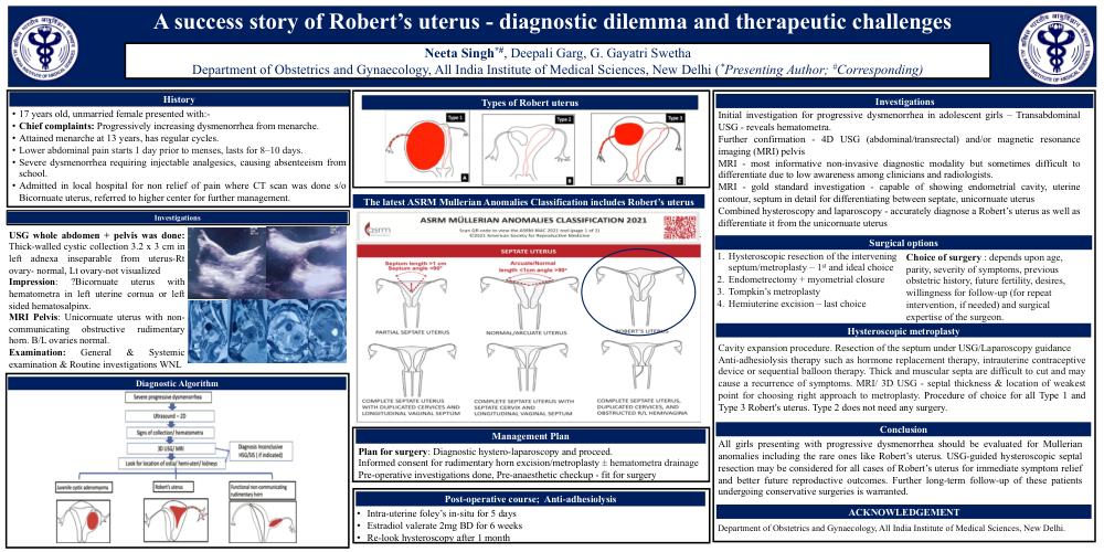
Title: “Diagnostic & Therapeutic challenges in managing a rare case of Robert’s Uterus”
Objective: To describe diagnostic dilemmas and management challenges of a rare Mullerian anomaly,” Robert Uterus”
Case report: A 17-year-old unmarried female presented with chief complaints of progressively increasing dysmenorrhoea since menarche at 13 years of agerequiring injectable analgesics and causing frequent absenteeism from school. A previous two-dimensional transabdominal ultrasound examination revealed presence of blood in the left side of the uterus without hematosalpinx or endometriosis which was confirmed on Three-dimensional (3D) ultrasound examination. Magnetic Resonance imaging (MRI) was suggestive ofuunicornuate uterus with non-communicating obstructive rudimentary horn with bilateral normal ovaries and kidneys. With a differential diagnosis of Juvenile cystic adenomyoma, Robert’s uterus and unicornuate uterus with functional non‑communicating rudimentary horn, a plan for diagnostic hystero-laparoscopy with rudimentary horn excision or metroplasty was made and proceeded accordingly. Intra-operatively, a dimple at uterine funduswas visualized with a few endometriotic spots on the surface of uterus and anterior abdominal wall.Bilateral tubes and ovaries were normal. Onhysteroscopy, right ostia and fundus were visualized normally while a thick left lateral uterine wall could be seen. On transabdominal needle aspiration from the obstructed Rt side, chocolate colored thick collection aspirated furtheron transillumination, only right uterine cavity was illuminated so a diagnosis of Robert’s uterus was made.Myometrial septum between the blind and the communicating hemi cavities was incised and extended following which left uterine cavity and ostia were visualized and entire uterine cavity could betrans-illuminated. To prevent intrauterine adhesions, intrauterine foley wasleftin-situ for 5 days and Estradiol valerate 2mg twice daily for 6 weeks was given. Re-look hysteroscopy after 6weeks revealed a normal uterine cavity with small residual septa at the fundus.No further intervention was done as the patient became asymptomatic.
Discussion:Only a few cases of Robert’s uterus are reported in the literature as awareness about the entity is poor among radiologists and clinicians leading to misdiagnosis and inappropriate surgeries. Combined hysteroscopy and laparoscopy can accurately diagnose a Robert’s uterus as well as differentiate it from unicornuate uterus or juvenile cystic adenomyoma. Choice of surgerydepends upon age, parity, severity of symptoms, previous obstetric history, future fertilityand surgical expertise of the surgeon.Hysteroscopic resection of the intervening septum/metroplasty is the first and ideal choice.
Conclusions:All girls presenting with progressive dysmenorrhea should be evaluated for Mullerian anomalies including the rare ones like Robert’s uterus. Hysteroscopic septal resection should be considered for immediate symptom relief and better future reproductive outcomes. Further long-term follow-up of these patients undergoing conservative surgeries is warranted.