2,281 posters, 63 videos, 56 audios, 43 topics, 18 sessions, 6,174 authors, 2,323 institutions
ePostersLive by SciGen Technologies S.A. All rights reserved.
RCOG World Congress 2025
June 23 - 25, 2025 | ExCeL London, UK
PP.0098
"Silent Time Bomb: A Case Report of a Ruptured Splenic Ectopic Pregnancy"
Poster Presenter
Authors
Affiliations
Part of Topic
Early pregnancy complications/early pregnancy and acute gynaecology
"Silent Time Bomb: A Case Report of a Ruptured Splenic Ectopic Pregnancy"
Case Report:
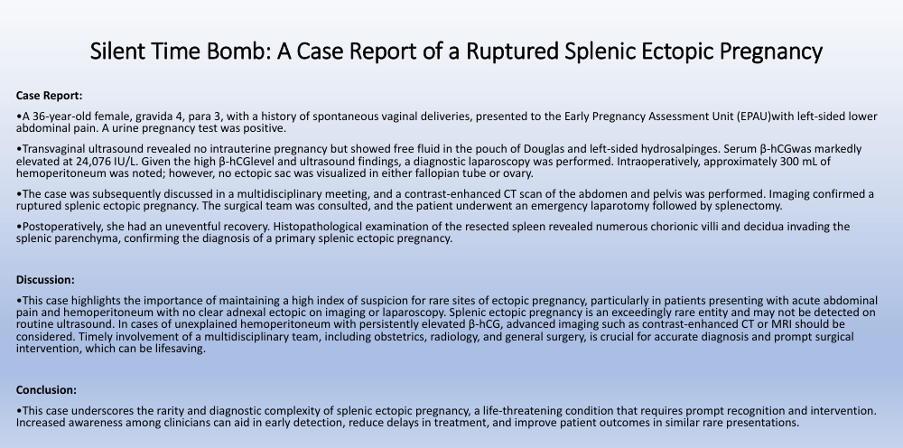
A 36-year-old female, gravida 4, para 3, with a history of spontaneous vaginal deliveries, presented to the Early Pregnancy Assessment Unit (EPAU) with left-sided lower abdominal pain. A urine pregnancy test was positive.
Transvaginal ultrasound revealed no intrauterine pregnancy but showed free fluid in the pouch of Douglas and left sided hydrosalpinges. Serum β-hCG was markedly elevated at 24,076 IU/L. Given the high β-hCG level and ultrasound findings, a diagnostic laparoscopy was performed. Intraoperatively, approximately 300 mL of hemoperitoneum was noted; however, no ectopic sac was visualized in either fallopian tube or ovary.
The case was subsequently discussed in a multidisciplinary meeting, and a contrast-enhanced CT scan of the abdomen and pelvis was performed. Imaging confirmed a ruptured splenic ectopic pregnancy. The surgical team was consulted, and the patient underwent an emergency laparotomy followed by splenectomy.
Postoperatively, she had an uneventful recovery. Histopathological examination of the resected spleen revealed numerous chorionic villi and decidua invading the splenic parenchyma, confirming the diagnosis of a primary splenic ectopic pregnancy.
Discussion:
This case highlights the importance of maintaining a high index of suspicion for rare sites of ectopic pregnancy, particularly in patients presenting with acute abdominal pain and hemoperitoneum with no clear adnexal ectopic on imaging or laparoscopy. Splenic ectopic pregnancy is an exceedingly rare entity and may not be detected on routine ultrasound. In cases of unexplained hemoperitoneum with persistently elevated β-hCG, advanced imaging such as contrast-enhanced CT or MRI should be considered. Timely involvement of a multidisciplinary team, including obstetrics, radiology, and general surgery, is crucial for accurate diagnosis and prompt surgical intervention, which can be lifesaving.
Conclusion:
This case underscores the rarity and diagnostic complexity of splenic ectopic pregnancy, a life-threatening condition that requires prompt recognition and intervention. Increased awareness among clinicians can aid in early detection, reduce delays in treatment, and improve patient outcomes in similar rare presentations.