2,281 posters, 63 videos, 56 audios, 43 topics, 18 sessions, 6,174 authors, 2,323 institutions
ePostersLive by SciGen Technologies S.A. All rights reserved.
RCOG World Congress 2025
June 23 - 25, 2025 | ExCeL London, UK
EP.0237
A case report of choriocarcinoma in unscarred uterus leading to perforated uterus
Part of Topic
Early pregnancy complications/early pregnancy and acute gynaecology
A case report : choriocarcinoma in unscarred uterus leading to perforated uterus
Authors: First Author and Main contributor Dr Asma Sadaqat, Second Author, Dr Aqsa Mandvia,Third Author, Dr Krishi Gowdra Revannasiddappa
Background
Gestational trophoblastic disease (GTD) encompasses a spectrum of conditions with varying levels of malignancy. Among these, choriocarcinoma is the most aggressive variant due to its rapid growth and high potential for spreading to other parts of the body. The incidence of choriocarcinoma is relatively rare, occurring in only two out of every 100,000 pregnancies.
Objective
This report focuses on a rare case where choriocarcinoma presented with acute emergency resulting in uterine perforation. The main aim is to bring in notice acute emergency cause in patients having history of chemotherapy followed by molar pregnancy so that these rarities cannot be missed.
Case Report
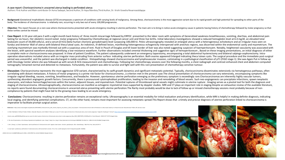
A 33-year-old para 3 with a eight-month back history of three month miscarriage followed by ERPOC presented to the labour room with symptoms of Generalized weakness breathlessness, vomiting, diarrhea, and abdominal pain lasting for six hours. Her obstetric record noted molar pregnancy Followed by Chemotherapy at regional cancer unit and three live births. Initial laboratory investigations showed a reduced hemoglobin level at 6.4 mg/dl, an elevated total leukocyte count of 34,000 cells/ul, and a serum β-hCG (beta-human chorionic gonadotropin) level surpassing 100,000 IU. Pelvic ultrasonography revealed an enlarged uterus with a heterogeneous echotexture and an irregular mass seen on fundus and Anterior Wall of uterus with bilateral theca luteal cysts. An indistinct, ill-defined lesion, manifesting heterogeneous echogenicity interspersed with anechoic regions, was discerned within the endometrial cavity and myometrium. The overlying myometrium was markedly thinned out with a suspicious area of rent. Fluid in Pouch of Douglas and till lower border of liver was also noted suggesting suspicion of haemperitonuem. Notably, heightened vascularity was associated with the lesion, with spectral waveform analyses identifying a low-resistance waveform. Further observations included omental thickening and suggestive signs of hemoperitoneum . Based on these imaging presentations, an initial diagnosis of either a complete invasive mole or choriocarcinoma was proposed. In View of patient Condition, The patient subsequently underwent an emergency laparotomy, encompassing a total abdominal hysterectomy and bilateral salpingo-oophorectomy. Intra-operative observations included uterus with an irregular mass protruding from anterior surface indicating uterine perforation. Both ovaries appeared Enlarged indicating theca lutean cysts. The surgical procedure and the immediate post-surgical period was uneventful, and the patient was discharged in stable condition . Histopathology showed choriocarcinoma and lymphovascular invasion, culminating in a pathological classification of pT1 (FIGO stage 1) .She was Again Put in follow up with Oncology Center where she was followed up with serum β-hCG measurement and chemotherapy. Following her chemotherapy sessions over the following months, a chest radiograph and contrast-enhanced chest and abdomen computed tomography remained normal and detected no metastasis. Fortunately, the patient was able to survive and fight well with this rare presentation of chemotherapy sensitive tumour.
Discussion
Choriocarcinoma represents the most aggressive GTD variant, characterized by its swift growth dynamics and significant metastatic potential. Typically, choriocarcinoma disseminates extensively via hematogenous pathways, often correlating with distant metastases. A history of molar pregnancy is a prime risk factor for choriocarcinoma , a criterion met in the present case.
The clinical presentation of choriocarcinoma can vary extensively, encompassing symptoms like irregular vaginal bleeding, nausea, vomiting, breathlessness, and headache. However, spontaneous uterine perforation emerging as the preliminary symptom is exceedingly rare.
Choriocarcinomas are inherently highly vascular tumors, synthesizing abundant angiogenic growth factors. Concurrently, there is pronounced cytotrophoblast proliferation, leading to the invasion and remodeling of blood vessels. Such neo-angiogenesis can precipitate infarctions, spawn neoplastic aneurysms, and induce hemorrhage/necrosis within the tumor, adjacent tissues, and myometrium. Potential ruptures of thrombosed spiral vessels might culminate in uterine rupture .
Imaging serves as a pivotal tool for both the diagnosis and staging of choriocarcinoma. Ultrasonographically, choriocarcinoma can manifest as echogenic myometrial mass supported by dopppler studies. MRI and CT plays an important role in staging.
Despite an exhaustive review of the available literature, no reports were found documenting choriocarcinoma in unscarred uterus presenting with uterine perforation.The Rarity most probably would be due to lack of follow up or missed chemotherapy sessions most probably because of non- compliance by patients that might have led to the growing mass leading to an acute emergency.
Conclusions
Choriocarcinoma resulting in uterine perforation remains an exceptional rarity. Ultrasonography is an essential modality for initial evaluation and primary identification, while MRI is helpful in making definite diagnosis, indicating local staging, and identifying potential complications. CT, on the other hand, remains most important for assessing metastatic spread.This Report shows that a timely and precise diagnosis of uterine perforation linked to choriocarcinoma is imperative to facilitate prompt surgical action.
References:
1.https://www.researchgate.net/publication/283236490_Choriocarcinoma_with_Uterine_Rupture_and_Shock_A_Rare_Case_Report
2. Choriocarcinoma-Driven Uterine Perforation in a Bicornuate Anatomy: An Elusive Encounter Maoulik Kumar P Modi 1,✉, Garima A Jasani 2, Nitin S Raithatha 3, Pokhraj P Suthar 4 Editors: Alexander Muacevic, John R Adler
3. Follow up to a case of ‘A silent rupture of extra horn of bicornuate uterus, due to molar pregnancy’. Singhai N, Chandrayan P, Verma P. https://www.worldwidejournals.com/international-journal-of-scientific-research-(IJSR)/recent_issues_pdf/2020/May/follow-up-to-a-case-of-a-silent-rupture-of-extra-horn-of-bicornuate-uterus-due-to-molar-pregnancy_May_2020_1588753978_3130258.pdf Int J Scient Research. 2020;9:2277–8179. [Google Scholar]
4. The role of hysterectomy in the therapy of gestational trophoblastic tumor. Xiang Y, Yang X, Du J, Song H. https://pubmed.ncbi.nlm.nih.gov/12899400/ Chin Med Sci J. 2000;15:45–48. [PubMed] [Google Scholar]
5. Xie C, Zheng L, Li Z, Zhao X. Spontaneous uterine perforation of choriocarcinoma with negative beta-human chorionic gonadotropin after chemotherapy. Med Princ Pract. 2011;20:570-73.
6. Case report: spontaneous perforation of a bicornuate uterus with concomitant sarcoma. Yim S, Yeo I, Lee M, Kyeong KS, Cho HY, Kang JB, Kyung MS. F1000Res. 2020;9:1143. doi: 10.12688/f1000research.25961.1. [DOI] [PMC free article] [PubMed] [Google Scholar]