2,281 posters, 63 videos, 56 audios, 43 topics, 18 sessions, 6,174 authors, 2,323 institutions
ePostersLive by SciGen Technologies S.A. All rights reserved.
RCOG World Congress 2025
June 23 - 25, 2025 | ExCeL London, UK
EP.0224
Acute abdomen from ruptured corpus luteal cyst
Poster Presenter
Part of Topic
Early pregnancy complications/early pregnancy and acute gynaecology
Introduction
Corpus luteum cyst is a functional cyst which is formed in the second phase of ovarian cycle. The natural history typically includes regression in the absence of pregnancy or regression after the first trimester of pregnancy and maturation of placenta. It is highly vascular structure and occasionally a subject of rupture. Blood loss is usually self-limited, but rarely can lead to massive hematoperitoneum and even death. Due to variable clinical presentation and sonographic appearance, the potential for misdiagnosis is high.
We present the case of a woman who underwent surgery with a preoperative diagnosis of ectopic pregnancy which was intra-operatively diagnosed as ruptured corpus luteal cyst causing the acute abdomen, this case is brought to report in view of rarity of its occurrence.
Case Report
40-year-old woman, G2P1, presented with lower abdominal pain and minimal vaginal bleeding at 6 weeks gestation. She was hemodynamically stable with a β-hCG level of 930.40 mIU/ml, admitted for observation as suspected ectopic pregnancy.
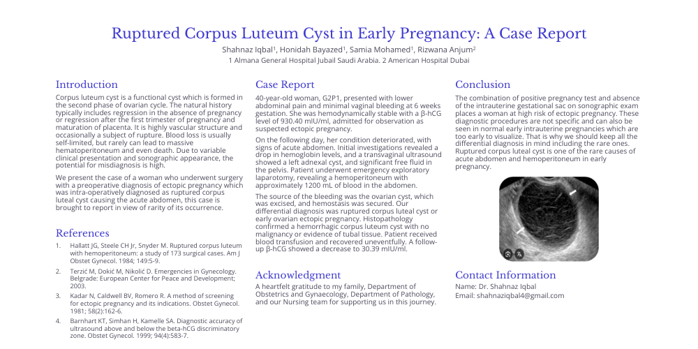
On the following day, her condition deteriorated, with signs of acute abdomen. Initial investigations revealed a drop in hemoglobin levels, and a transvaginal ultrasound showed a left adnexal cyst, and significant free fluid in the pelvis. Patient underwent emergency exploratory laparotomy, revealing a hemoperitoneum with approximately 1200 mL of blood in the abdomen.
The source of the bleeding was the ovarian cyst, which was excised, and hemostasis was secured. Our differential diagnosis was ruptured corpus luteal cyst or early ovarian ectopic pregnancy. Histopathology confirmed a hemorrhagic corpus luteum cyst with no malignancy or evidence of tubal tissue. Patient received blood transfusion and recovered uneventfully. A follow-up β-hCG showed a decrease to 30.39 mIU/ml.
Conclusion
The combination of positive pregnancy test and absence of the intrauterine gestational sac on sonographic exam places a woman at high risk of ectopic pregnancy. These diagnostic procedures are not specific and can also be seen in normal early intrauterine pregnancies which are too early to visualize. That is why we should keep all the differential diagnosis in mind including the rare ones. Ruptured corpus luteal cyst is one of the rare causes of acute abdomen and hemoperitoneum in early pregnancy.
References
A heartfelt gratitude to my family, Department of Obstetrics and Gynaecology, Department of Pathology, and our Nursing team for supporting us in this journey.
Contact Information
Name: Dr. Shahnaz Iqbal
Email: shahnaziqbal4@gmail.com