2,281 posters, 63 videos, 56 audios, 43 topics, 18 sessions, 6,174 authors, 2,323 institutions
ePostersLive by SciGen Technologies S.A. All rights reserved.
RCOG World Congress 2025
June 23 - 25, 2025 | ExCeL London, UK
EP.0223
A case of a rudimentary horn ectopic pregnancy diagnosed and managed laparoscopically
Poster Presenter
Authors
Affiliations
Part of Topic
Early pregnancy complications/early pregnancy and acute gynaecology
A case of a rudimentary horn ectopic pregnancy diagnosed and managed laparoscopically
Dr Somia Khalid (ST7 Obstetrics and gynaecology), Dr Nilofar Noor (ST7 Obstetrics and gynaecology) Mr Jaydip Dasgupta (Consultant Gynaecologist)
Royal Derby Hospital, Derby, United Kingdom
A 29-year-old female presented at approximately eight weeks gestation with a two-day history light vaginal bleeding and mild left sided pain. This was her third pregnancy, following two previous caesarean sections. She was assessed in the Emergency department and deemed clinically stable therefore an appointment was booked with the Early pregnancy unit for the following day. Her serum BHCG was 5413. A pelvic ultrasound scan was performed.
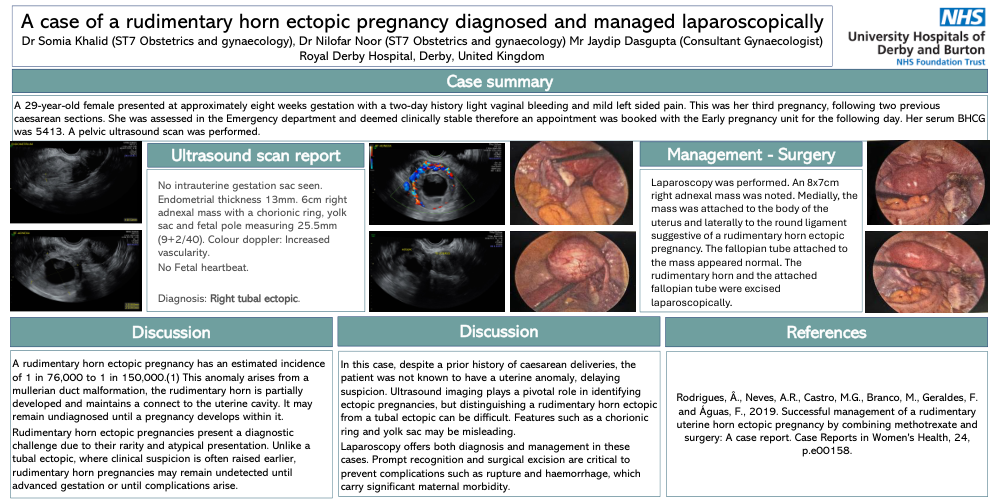
No intrauterine gestation sac seen. Endometrial thickness 13mm. 6cm right adnexal mass with a chorionic ring, yolk sac and fetal pole measuring 25.5mm (9+2/40). Colour doppler: Increased vascularity.
No Fetal heartbeat.
Diagnosis: Right tubal ectopic.
A rudimentary horn ectopic pregnancy has an estimated incidence of 1 in 76,000 to 1 in 150,000.(1) This anomaly arises from a mullerian duct malformation, the rudimentary horn is partially developed and maintains a connect to the uterine cavity. It may remain undiagnosed until a pregnancy develops within it.
Rudimentary horn ectopic pregnancies present a diagnostic challenge due to their rarity and atypical presentation. Unlike a tubal ectopic, where clinical suspicion is often raised earlier, rudimentary horn pregnancies may remain undetected until advanced gestation or until complications arise.
In this case, despite a prior history of caesarean deliveries, the patient was not known to have a uterine anomaly, delaying suspicion. Ultrasound imaging plays a pivotal role in identifying ectopic pregnancies, but distinguishing a rudimentary horn ectopic from a tubal ectopic can be difficult. Features such as a chorionic ring and yolk sac may be misleading.
Laparoscopy offers both diagnosis and management in these cases. Prompt recognition and surgical excision are critical to prevent complications such as rupture and haemorrhage, which carry significant maternal morbidity.
Laparoscopy was performed. An 8x7cm right adnexal mass was noted. Medially, the mass was attached to the body of the uterus and laterally to the round ligament suggestive of a rudimentary horn ectopic pregnancy. The fallopian tube attached to the mass appeared normal. The rudimentary horn and the attached fallopian tube were excised laparoscopically.