2,281 posters, 63 videos, 56 audios, 43 topics, 18 sessions, 6,174 authors, 2,323 institutions
ePostersLive by SciGen Technologies S.A. All rights reserved.
RCOG World Congress 2025
June 23 - 25, 2025 | ExCeL London, UK
EP.2229
VIN-teresting Findings: Two cases of vulval intraepithelial neoplasia
Poster Presenter
Part of Topic
Vulval disease
VIN-teresting Findings: Two cases of vulval intraepithelial neoplasia
Dr Jennifer Simpson, Dr Jenny Chung , Miss Elaine Palmer
Dr Randa Akel, Dr Jacqueline McDermott, Miss Diana Marcus , Miss Helen Staley
Chelsea and Westminster Foundation Trust
Human papillomavirus (HPV) independent vulval intraepithelial neoplasia (VIN) can rapidly progress to vulval squamous cell carcinoma (VSCC) when left untreated. The different types of HPV-independent VIN include;
We present two cases of dVIN which were not straightforward to diagnose clinically.
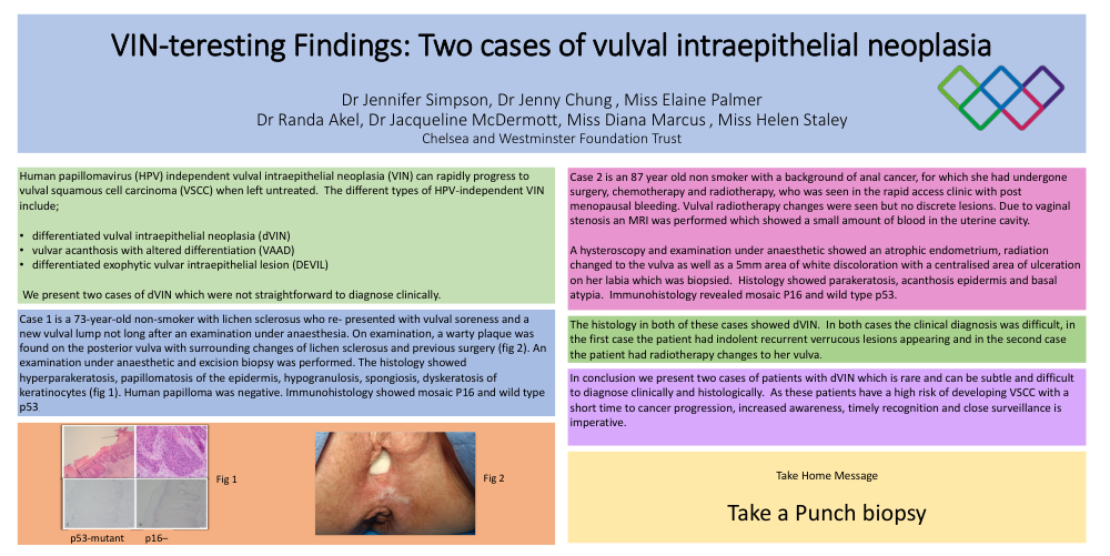
Case 1 is a 73-year-old non-smoker with lichen sclerosus who re- presented with vulval soreness and a new vulval lump not long after an examination under anaesthesia. On examination, a warty plaque was found on the posterior vulva with surrounding changes of lichen sclerosus and previous surgery (fig 2). An examination under anaesthetic and excision biopsy was performed. The histology showed hyperparakeratosis, papillomatosis of the epidermis, hypogranulosis, spongiosis, dyskeratosis of keratinocytes (fig 1). Human papilloma was negative. Immunohistology showed mosaic P16 and wild type p53.
Case 2 is an 87 year old non smoker with a background of anal cancer, for which she had undergone surgery, chemotherapy and radiotherapy, who was seen in the rapid access clinic with post menopausal bleeding. Vulval radiotherapy changes were seen but no discrete lesions. Due to vaginal stenosis an MRI was performed which showed a small amount of blood in the uterine cavity.
A hysteroscopy and examination under anaesthetic showed an atrophic endometrium, radiation changed to the vulva as well as a 5mm area of white discoloration with a centralised area of ulceration on her labia which was biopsied. Histology showed parakeratosis, acanthosis epidermis and basal atypia. Immunohistology revealed mosaic P16 and wild type p53
The histology in both of these cases showed dVIN. In both cases the clinical diagnosis was difficult, in the first case the patient had indolent recurrent verrucous lesions appearing and in the second case the patient had radiotherapy changes to her vulva.
In conclusion we present two cases of patients with dVIN which is rare and can be subtle and difficult to diagnose clinically and histologically. As these patients have a high risk of developing VSCC with a short time to cancer progression, increased awareness, timely recognition and close surveillance is imperative.