2,281 posters, 63 videos, 56 audios, 43 topics, 18 sessions, 6,174 authors, 2,323 institutions
ePostersLive by SciGen Technologies S.A. All rights reserved.
RCOG World Congress 2025
June 23 - 25, 2025 | ExCeL London, UK
EP.0220
Crucial role of urinary pregnancy test in reproductive women with abdominal pain
Poster Presenter
Authors
Affiliations
Part of Topic
Early pregnancy complications/early pregnancy and acute gynaecology
Title: Crucial Role of Urinary Pregnancy Test in Reproductive Women with Abdominal Pain
Objective: To emphasize the importance of urine pregnancy tests in reproductive women and interprofessional communication.
Case Report:
Clinical History:
A 28-year-old female presented to the Emergency Department with right iliac fossa (RIF) pain. Her last menstrual period (LMP) was four weeks prior, and she had experienced vaginal spotting since that period. Despite the pain, she remained hemodynamically stable upon presentation.
Initial Evaluation:
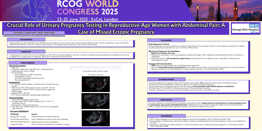
On clinical examination, there was mild tenderness in both the suprapubic region and the right iliac fossa. Laboratory investigations revealed a haemoglobin concentration of 132 g/L (Fig1). A urine dipstick test demonstrated significant hematuria (erythrocytes 4+), prompting consideration of underlying urinary tract pathology.
Radiological Assessment:
In order to exclude renal pathology, including urolithiasis, a computed tomography (CT) scan of the urinary tract was performed. The CT scan revealed normal bilateral kidneys; however, a pelvic hematoma was identified, which was interpreted as being either within the bladder region or as a soft tissue mass within the uterus. Additionally, mild to moderate abdomino-pelvic ascites was noted. In response to these findings, a serum beta-human chorionic gonadotropin (BhCG) test was ordered, along with a pelvic ultrasonography (US).
Ultrasonographic Findings:
The pelvic ultrasound demonstrated a heterogeneous mass with approximate dimensions of 81 mm x 41 mm. The differential diagnosis included an ectopic pregnancy or ovarian torsion (Fig 2a and 2b). Adjacent to this mass, a 56 mm fluid collection exhibiting low-level echoes was identified, suggestive of pelvic haemorrhage (Fig 3). With these findings, the patient was promptly referred to the on-call gynaecological team. Owing to the pending serum β-hCG results and the absence of pre-existing documentation of a urine pregnancy test, a repeat urine pregnancy test was performed, yielding a positive result. The subsequent BhCG level was confirmed at 237 IU/L.
Surgical Intervention and Intraoperative Findings:
Given the clinical and radiological evidence, the patient was taken to the operating theatre. Intraoperatively, approximately 1.7 liters of hemoperitoneum were evacuated. Further surgical exploration identified a right-sided ectopic pregnancy measuring approximately 4 cm x 3 cm. A right salpingectomy was performed to manage the ectopic gestation.
Postoperative Management and Outcome:
Following the surgical intervention, the patient was managed postoperatively with close observation. She remained clinically stable, receiving supportive care including iron infusion to optimize her hemoglobin levels. After a 48-hour period of inpatient observation, the patient was debriefed and subsequently discharged in stable condition.
Introduction:
Definition and Overview
- Ectopic pregnancy is defined as the implantation of a fertilized ovum outside the uterine cavity.
- The most frequent site of implantation is within the fallopian tubes.
Incidence in the UK
- Occurs in approximately 1 in every 90 pregnancies.
- This equates to around 11,000 cases annually, highlighting its clinical significance.
Diagnostic Methods
- Clinical Assessment: Comprehensive evaluation including history and physical examination and urinary pregnancy test.
- Imaging Techniques:
- Transvaginal ultrasound (TVS) is the imaging modality of choice, permitting direct visualization of the gestational sac and precise localization.
- Biochemical Analysis:
- Measurements of human chorionic gonadotropin (hCG) help to plan the management of TVS-diagnosed ectopic pregnancy.
Importance of Early Diagnosis
- Early identification is critical to prevent life-threatening complications, such as tubal rupture and significant internal hemorrhage.
- Prompt diagnosis facilitates timely management, which may include medical treatment (e.g., methotrexate) or minimally invasive surgical interventions, thereby improving patient outcomes.
Rationale of the case report:
- This case underscores the significance of urinary pregnancy testing for all women of reproductive age presenting with abdominal pain.
- The dilemma of the diagnosis of internal bleeding due to compensatory mechanism of the body to blood loss.
- Emphasize the importance of proper documentation and communication.
Description:
Early detection is pivotal to reducing maternal mortality, morbidity, psychological trauma, and to enhancing future reproductive outcomes, while also shortening hospital stays and lowering treatment costs. Consequently, routine triage with a urine pregnancy test is essential for all reproductive-aged women to exclude early pregnancy-related complications. In this case, diagnostic delay occurred due to several factors: the presence of haematuria, incomplete documentation of a urine pregnancy test, and the patient’s maintained hemodynamic stability. Notably, a young, healthy woman can compensate for significant blood loss—up to 15% of her total blood volume—and the physiological increase in circulating volume during early pregnancy may further mask the clinical signs of hypovolemic shock. Despite these factors, the patient’s concerns regarding abdominal pain were appropriately addressed; imaging studies identified pelvic fluid, thereby prompting further pregnancy-related investigations. Subsequent management was timely and appropriate, resulting in the patient being discharged in a stable condition.
Learning Points
This case underscores the diagnostic dilemma posed by the patient's stable hemodynamic status and normal haemoglobin level, which could obscure the severity of the underlying condition. The lack of clear documentation of the urine pregnancy test further complicated the diagnostic process. Multi-modal imaging, including CT and ultrasound, plays a crucial role in differentiating pelvic pathology. Timely surgical intervention in suspected ruptured ectopic pregnancy is lifesaving and significantly improves patient outcomes.