2,281 posters, 63 videos, 56 audios, 43 topics, 18 sessions, 6,174 authors, 2,323 institutions
ePostersLive by SciGen Technologies S.A. All rights reserved.
RCOG World Congress 2025
June 23 - 25, 2025 | ExCeL London, UK
EP.0218
Decidualised Ovarian Endometrioma Mimicking Malignancy in Pregnancy
Poster Presenter
Authors
Affiliations
Part of Topic
Early pregnancy complications/early pregnancy and acute gynaecology
Title
Decidualised Ovarian Endometrioma Mimicking Malignancy in Pregnancy
Objective: This case report explores a rare presentation of decidualised ovarian endometrioma in a 38-year-old G2P0 patient at 13+3 weeks of pregnancy, creating a diagnostic dilemma. Decidualised endometriosis occurs when ectopic endometrial tissue undergoes a decidual reaction, triggered by the hormonal changes of pregnancy. Although rare, it can mimic malignancy, complicating diagnosis.
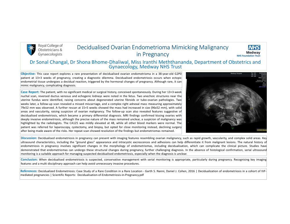
Case Report: The patient, with no significant medical or surgical history, conceived spontaneously. During her 13+3-week nuchal scan, reversed ductus venosus and echogenic kidneys were noted in the fetus. Two anechoic structures near the uterine fundus were identified, raising concerns about degenerated uterine fibroids or tubo-ovarian pathologies. Two weeks later, a follow-up scan revealed a missed miscarriage, and a complex right adnexal mass measuring approximately 79x52 mm was observed. A further rescan at 15+5 weeks showed the mass had increased in size (96x52 mm), with solid areas and vascularity, raising suspicion of ovarian malignancy. The follow-up scan also revealed features suggestive of decidualised endometriosis, which became a primary differential diagnosis. MRI findings confirmed kissing ovaries with deeply invasive endometriosis, although the precise nature of the mass remained unclear, a suspicion of malignancy was highlighted by the radiologists. The CA125 level was mildly elevated at 48, while all other blood markers were normal. The patient was referred for laparoscopy, cystectomy, and biopsy, but opted for close monitoring instead, declining surgery after being made aware of the risks. Her repeat scan showed resolution of the findings but endometriomas remained.
Discussion: Decidualised endometriosis in pregnancy can present with imaging features resembling ovarian malignancy, such as rapid growth, vascularity, and complex solid areas. Key ultrasound characteristics, including the “ground glass” appearance and intracystic excrescences and adhesions can help differentiate it from malignant lesions. The natural history of endometriosis in pregnancy involves significant changes in the morphology of endometriomas, including decidualisation, which can complicate the clinical picture. Studies have demonstrated that endometriomas can undergo these structural changes during pregnancy, further challenging diagnosis. In the absence of histological confirmation, serial ultrasound monitoring is a suitable approach for managing suspected decidualised endometriosis, especially when the diagnosis is unclear.
Conclusion: When decidualised endometriosis is suspected, conservative management with serial monitoring is appropriate, particularly during pregnancy. Recognising key imaging features and a multi disciplinary approach can help avoid unnecessary invasive procedures.