2,281 posters, 63 videos, 56 audios, 43 topics, 18 sessions, 6,174 authors, 2,323 institutions
ePostersLive by SciGen Technologies S.A. All rights reserved.
RCOG World Congress 2025
June 23 - 25, 2025 | ExCeL London, UK
EP.0214
Ovarian Ectopic Pregnancy in a patient with IUD in situ: Case report.
Part of Topic
Early pregnancy complications/early pregnancy and acute gynaecology
Ovarian Ectopic Pregnancy in a patient with IUD in situ: Case report.
Maria Camila Cetina Grajales, MD1 , Kelly Moralba Agudelo Cañón, MD 2 , Ana Gabriela Martínez Charry. MD 3 , Andrea Paola Plazas Bedoya, MD 4.
1Pontificia Universidad Javeriana, Bogotá, Colombia; 2Pontificia Universidad Javeriana, Bogotá, Colombia; 3Pontificia Universidad Javeriana, Bogotá, Colombia; 4Pontificia Universidad Javeriana, Bogotá, Colombia.
INTRODUCTION
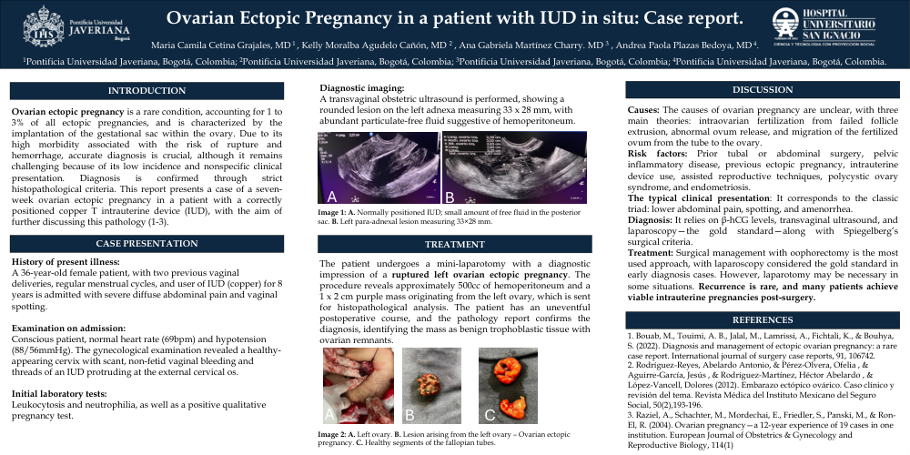
Ovarian ectopic pregnancy is a rare condition, accounting for 1 to 3% of all ectopic pregnancies, and is characterized by the implantation of the gestational sac within the ovary. Due to its high morbidity associated with the risk of rupture and hemorrhage, accurate diagnosis is crucial, although it remains challenging because of its low incidence and nonspecific clinical presentation. Diagnosis is confirmed through strict histopathological criteria. This report presents a case of a seven-week ovarian ectopic pregnancy in a patient with a correctly positioned copper T intrauterine device (IUD), with the aim of further discussing this pathology (1-3).
CASE PRESENTATION
History of present illness:
A 36-year-old female patient, with two previous vaginal deliveries, regular menstrual cycles, and user of IUD (copper) for 8 years is admitted with severe diffuse abdominal pain and vaginal spotting.
Examination on admission:
Conscious patient, normal heart rate (69bpm) and hypotension (88/56mmHg). The gynecological examination revealed a healthy-appearing cervix with scant, non-fetid vaginal bleeding and threads of an IUD protruding at the external cervical os.
Initial laboratory tests:
Leukocytosis and neutrophilia, as well as a positive qualitative pregnancy test.
Diagnostic imaging:
A transvaginal obstetric ultrasound is performed, showing a rounded lesion on the left adnexa measuring 33 x 28 mm, with abundant particulate-free fluid suggestive of hemoperitoneum.
Image 1: A. Normally positioned IUD; small amount of free fluid in the posterior sac.B. Left para-adnexal lesion measuring 33×28 mm.
TREATMENT
The patient undergoes a mini-laparotomy with a diagnostic impression of a ruptured left ovarian ectopic pregnancy. The procedure reveals approximately 500cc of hemoperitoneum and a 1 x 2 cm purple mass originating from the left ovary, which is sent for histopathological analysis. The patient has an uneventful postoperative course, and the pathology report confirms the diagnosis, identifying the mass as benign trophoblastic tissue with ovarian remnants.
Image 2: A. Left ovary. B. Lesion arising from the left ovary – Ovarian ectopic pregnancy. C. Healthy segments of the fallopian tubes.
DISCUSSION
Causes: The causes of ovarian pregnancy are unclear, with three main theories: intraovarian fertilization from failed follicle extrusion, abnormal ovum release, and migration of the fertilized ovum from the tube to the ovary. (1)
Risk factors: Prior tubal or abdominal surgery, pelvic inflammatory disease, previous ectopic pregnancy, intrauterine device use, assisted reproductive techniques, polycystic ovary syndrome, and endometriosis. (3)
The typical clinical presentation: It corresponds to the classic triad: lower abdominal pain, spotting, and amenorrhea.
Diagnosis: It relies on β-hCG levels, transvaginal ultrasound, and laparoscopy—the gold standard—along with Spiegelberg’s surgical criteria. (5)
Treatment: Surgical management with oophorectomy is the most used approach, with laparoscopy considered the gold standard in early diagnosis cases. However, laparotomy may be necessary in some situations. Recurrence is rare, and many patients achieve viable intrauterine pregnancies post-surgery.