2,281 posters, 63 videos, 56 audios, 43 topics, 18 sessions, 6,174 authors, 2,323 institutions
ePostersLive by SciGen Technologies S.A. All rights reserved.
RCOG World Congress 2025
June 23 - 25, 2025 | ExCeL London, UK
EP.0202
Enhanced Myometrial Vascularity: Can expectant management be first-line despite marked high-velocity circulation?
Poster Presenter
Authors
Affiliations
Part of Topic
Early pregnancy complications/early pregnancy and acute gynaecology
Enhanced Myometrial Vascularity: Can expectant management be first-line despite marked high-velocity circulation? ]
Introduction
• Enhanced myometrial vascularity (EMV) is a rare phenomenon of vascular lesions involving the myometrium. It is a physiological response to retained pregnancy tissue after birth or following miscarriage or termination of pregnancy (RPOC). • EMV are characterised by high-velocity circulation within the myometrium due to the direct communication between the high pressure arterial system and the low-pressure venous system, as seen in the spiral/ radial arteries within the intervillous spaces of the placental bed. • It should not be confused with uterine arteriovenous malformations (AVM) as EMV only occur after recent pregnancy- failure of subinvolution of the myometrial placental bed due to the persistence of retained tissue. • True congenital AVM are rare. The incidence of EMV is uncertain due to the interchanged terminology use of EMV and AVM in literature, the reported EMV prevalence is as much as 1.5-6.3% • Diagnosis is made by transvaginal-ultrasound (TVUS): tubular, tortuous, anechoic structures in the myometrium on greyscale sonography. Doppler assessment shows unusually high flow velocities, beneath the RPOC which may or may not be preceded by major haemorrhage. Therapeutic dilemmas arise if pre emptive radiological intervention is considered when marked Doppler vascularity is seen and major bleeding is anticipated . • Resolution always occurs once the tissue is removed and EMV vascularity does not appear to correlate with surgical blood loss. Also, the amount of residual tissue seen with EMV is sometimes limited or undetectable on TVUS and only seen at hysteroscopy. • We present the management of EMV diagnosed in the presence of RPOC after Surgical Management of Miscarriage.
Case Report
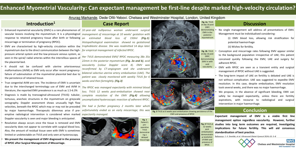
32-year-old nulliparous woman underwent surgical management of miscarriage at 10 weeks’ gestation with an estimated blood loss of 150ml (Fig.1). Histopathological examination showed no gestational trophoblastic disease. She was readmitted 14 days later for empirical management of infected RPOC. Her TVUS demonstrated EMV/ RPOC measuring 38x 35x 22mm in the posterior myometrium (Fig. 2a and b), avid vascularity (colour Doppler score 4). EMV was reconfirmed by CT-angiogram and she underwent bilateral selective uterine artery embolisation (UAE). The patient was closely monitored with weekly TVUS for 6 weeks and then fortnightly. (Fig.3) The RPOC was managed expectantly with minimal blood loss. TVUS 12 weeks post-embolisation showed near complete resolution of the EMV (Fig.4) allowing uncomplicated hysteroscopic resection of adherent RPOC. She had a further pregnancy 2 months later which unfortunately ended as an early miscarriage; this was expectantly managed
Discussion
• No single management will address all presentations of EMV. Management must be individualised considering: (i) EMV blood loss, allowing risk stratification for potential haemorrhage (ii) Wishes for fertility. • Conception and miscarriage rates following EMV appear similar to the background population irrespective of UAE; this patient conceived quickly following the EMV, UAE and surgery for adherent RPOC. • EMV with RPOC are seen as a transient entity and surgical management of RPOC without UAE is suggested. • The long-term impact of UAE on fertility is debated and UAE is not without complication. UAE was suggested to expedite EMV resolution. In this case, despite embolization, EMV resolution took several weeks, and there was no major haemorrhage. • We propose, in the absence of significant bleeding, EMV can safely be managed expectantly, unless there are fertility aspirations, with recourse to radiological and surgical intervention in major haemorrhage.
Conclusion
Expectant management of EMV is a viable first line management option regardless vascularity. However, further studies into long term outcomes are required, investigating implications for future fertility. This will aid consensus standardisation of best practice.
Reference: 1. K. Dewilde, et al. Ultrasound Obstet Gynecol 2024; 63: 5–