2,281 posters, 63 videos, 56 audios, 43 topics, 18 sessions, 6,174 authors, 2,323 institutions
ePostersLive by SciGen Technologies S.A. All rights reserved.
RCOG World Congress 2025
June 23 - 25, 2025 | ExCeL London, UK
EP.0199
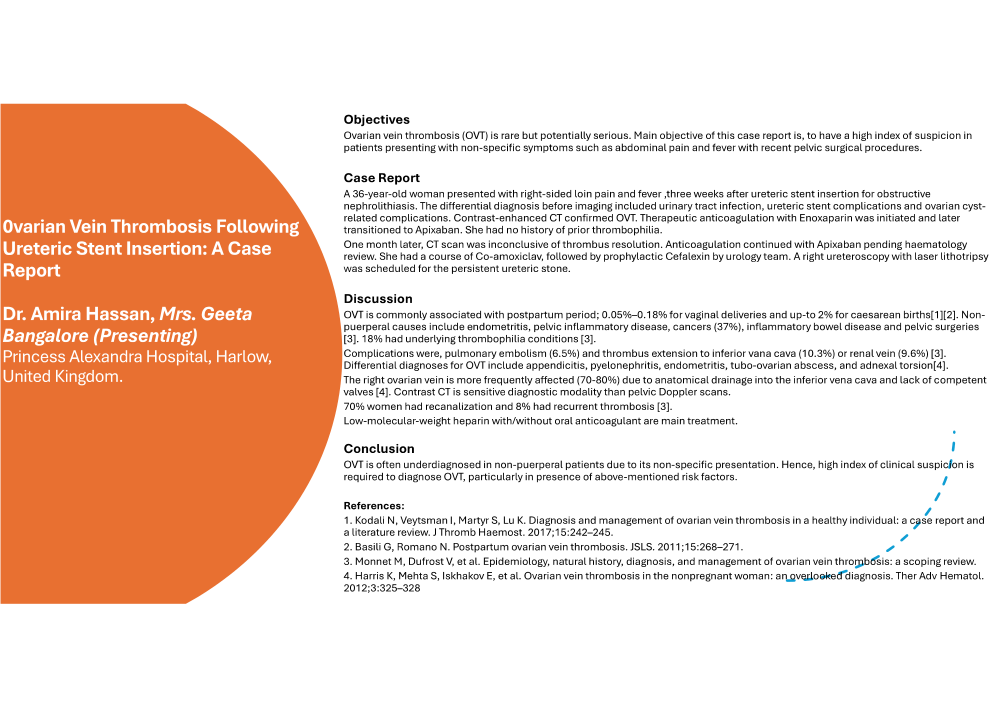
Ovarian Vein Thrombosis Following Ureteric Stent Insertion: A Case Report
Poster Presenter
Authors
Affiliations
Part of Topic
Early pregnancy complications/early pregnancy and acute gynaecology
Ovarian Vein Thrombosis Following Ureteric Stent Insertion: A Case Report
Objectives
Ovarian vein thrombosis (OVT) is rare but potentially serious. Main objective of this case report is, to have a high index of suspicion in patients presenting with non-specific symptoms such as abdominal pain and fever with recent pelvic surgical procedures.
Case Report
A 36-year-old woman presented with right-sided loin pain and fever ,three weeks after ureteric stent insertion for obstructive nephrolithiasis. The differential diagnosis before imaging included urinary tract infection, ureteric stent complications and ovarian cyst-related complications. Contrast-enhanced CT confirmed OVT. Therapeutic anticoagulation with Enoxaparin was initiated and later transitioned to Apixaban. She had no history of prior thrombophilia.
One month later, CT scan was inconclusive of thrombus resolution. Anticoagulation continued with Apixaban pending haematology review. She had a course of Co-amoxiclav, followed by prophylactic Cefalexin by urology team. A right ureteroscopy with laser lithotripsy was scheduled for the persistent ureteric stone.
Discussion
OVT is commonly associated with postpartum period; 0.05%–0.18% for vaginal deliveries and up-to 2% for caesarean births[1][2]. Non-puerperal causes include endometritis, pelvic inflammatory disease, cancers (37%), inflammatory bowel disease and pelvic surgeries [3]. 18% had underlying thrombophilia conditions [3].
Complications were, pulmonary embolism (6.5%) and thrombus extension to inferior vana cava (10.3%) or renal vein (9.6%) [3]. Differential diagnoses for OVT include appendicitis, pyelonephritis, endometritis, tubo-ovarian abscess, and adnexal torsion[4].
The right ovarian vein is more frequently affected (70-80%) due to anatomical drainage into the inferior vena cava and lack of competent valves [4]. Contrast CT is sensitive diagnostic modality than pelvic Doppler scans.
70% women had recanalization and 8% had recurrent thrombosis [3].
Low-molecular-weight heparin with/without oral anticoagulant are main treatment.
Conclusion
OVT is often underdiagnosed in non-puerperal patients due to its non-specific presentation. Hence, high index of clinical suspicion is required to diagnose OVT, particularly in presence of above mentioned risk factors.
References:
1. Kodali N, Veytsman I, Martyr S, Lu K. Diagnosis and management of ovarian vein thrombosis in a healthy individual: a case report and a literature review. J Thromb Haemost. 2017;15:242–245.
2. Basili G, Romano N. Postpartum ovarian vein thrombosis. JSLS. 2011;15:268–271.
3. Monnet M, Dufrost V, et al. Epidemiology, natural history, diagnosis, and management of ovarian vein thrombosis: a scoping review.
4. Harris K, Mehta S, Iskhakov E, et al. Ovarian vein thrombosis in the nonpregnant woman: an overlooked diagnosis. Ther Adv Hematol. 2012;3:325–328.