2,281 posters, 63 videos, 56 audios, 43 topics, 18 sessions, 6,174 authors, 2,323 institutions
ePostersLive by SciGen Technologies S.A. All rights reserved.
RCOG World Congress 2025
June 23 - 25, 2025 | ExCeL London, UK
EP.0198
Uterine Rupture at 31 weeks’ Gestation of an Intramural Pregnancy following Myomectomy
Poster Presenter
Authors
Gabriel Awadzi, Anahit Zakaryan, Hend Hadawi, Sukhy Bhullar, Mark Waterstone
Affiliations
Part of Topic
Early pregnancy complications/early pregnancy and acute gynaecology
Uterine Rupture at 31 weeks’ Gestation of an Intramural Pregnancy; A Salutary Lesson for us all
Gabriel Awadzi, Anahit Zakaryan, Hend Hadawi, Sukhy Bhullar, Mark Waterstone, Darent Valley Hospital, Dartford
Introduction
Intramural pregnancies are an exceedingly rare [1] form of ectopic pregnancy, where the embryo implants wholly within the myometrium away from the cavity or the Fallopian tube. Diagnosis of intramural implantation is particularly difficult. Previous authors [2] have reported on the role of ultrasound and MRI. The risk of life-threatening haemorrhage due to uterine rupture is significant and a hysterectomy is often required in such cases. The pathogenesis of intramural pregnancies is uncertain. Risk factors include history of uterine trauma, myomectomy, caesarean section or dilatation and curettage. Trauma results in the formation of a sinus tract within the endometrium, enabling the embryo to penetrate into the myometrium. Pegnancies rarely exceed 12 weeks’ gestation as the risk of uterine rupture and maternal mortality are substantial [3]. It is therefore remarkable that our patient’s pregnancy continued until 31 weeks’ gestation.
Case Report
A 33 year old woman, Para 0+1 (previous miscarriage) with a history of open myomectomy presented at 6+5 weeks’ gestation with pelvic pain and vaginal bleeding. A TV scan queried the location of her pregnancy, there appeared to be a thin rim of myometrium around the gestation sac. Subsequent scans did not show any abnormal findings. Throughout her pregnancy she had repeated episodes of unexplained abdominal pains.
At 17+5 weeks gestation, she was admitted with right iliac fossa pain. She was treated for constipation and discharged. Four days later, she re-presented with identical symptoms. Differential diagnoses included: constipation, red degeneration of fibroids, ovarian cyst accident and appendicitis. She wall. The impression was of a ruptured haemorrhagic ovarian cyst. She had further bouts of abdominal pain over the subsequent weeks. underwent an open appendicectomy. The appendix looked normal and approximately 300mls of dark red blood was seen in the pelvis. An attempt to visualise her pelvic organs was limited by the “grid iron” incision.
At 31 weeks’ gestation she presented with significant abdominal pain with suspected preterm labour. She had a normal CTG. Her pain worsened several hours later and she transferred to the operating theatre for a Category1 Caesarean section for a possible abruption. On entry into the abdomen, there was extensive haemoperitoneum seen with a uterine rupture. A fresh stillborn baby was seen in the epigastrium. The placenta was morbidly adherent and was removed with significant difficulty. Her blood loss was uncontrollable and a subtotal hysterectomy was performed by two consultants. Following massive transfusions and a brief spell in the ITU, she recovered well and was discharged home within a week.
Histology of the uterus confirmed several leiomyomata. There was extensive infiltration of the myometrium by trophoblast and chorionic villi without associated decidualised endometrium. There appeared to have been serosal rupture and the appearances were consistent with placenta percreta..
Discussion:
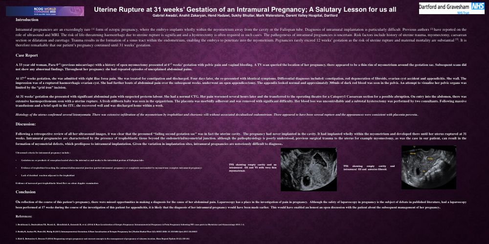
Following a retrospective review of all her ultrasound images, it was clear that the presumed “failing second gestation sac” was in fact the uterine cavity. The pregnancy had never implanted in the cavity. It had implanted wholly within the myometrium and developed there until her uterus ruptured at 31 weeks. Intramural pregnancies are characterised by the presence of trophoblastic tissue beyond the endometrial/myometrial junction. although the pathophysiology is poorly understood, previous surgical trauma to the uterus for example myomectomy, as was the case in our patient, can result in the formation of myometrial defects, which predispose to intramural implantation. Given the variation in implantation sites, intramural pregnancies are notoriously difficult to diagnose.