2,281 posters, 63 videos, 56 audios, 43 topics, 18 sessions, 6,174 authors, 2,323 institutions
ePostersLive by SciGen Technologies S.A. All rights reserved.
RCOG World Congress 2025
June 23 - 25, 2025 | ExCeL London, UK
EP.0194
Ruptured Cornual Pregnancy: A Rare and Challenging Obstetric Emergency
Poster Presenter
Authors
Affiliations
Part of Topic
Early pregnancy complications/early pregnancy and acute gynaecology
Ruptured Cornual Pregnancy: A Rare and Challenging Obstetric Emergency
Authors: A. Brennan, M. Abdelrahman, B.A. Muresan
Introduction:
Ectopic pregnancy (EP) occurs at a rate of 11 per 1,000 pregnancies. EP rupture remains the leading cause of maternal mortality during the first trimester and accounts for an incidence of 5%-10% of all pregnancy-related deaths (1).
Cornual pregnancies (CP) are a rare subtype of EP, accounting for 2-4% of ectopic pregnancies, occurring when the gestational sac implants in the interstitial portion of the fallopian tube. This approximately 1-2cm long region transverses the myometrium in the uterine fundus in a highly vascular area supplied by Sampson’s artery, connected to the uterine and ovarian arteries[2].
Cornual Ectopics carry a mortality rate of 2.0%–2.5%, 2 – 5 times higher than that of other forms of ectopic pregnancies[3]. Due to:
Case Report:
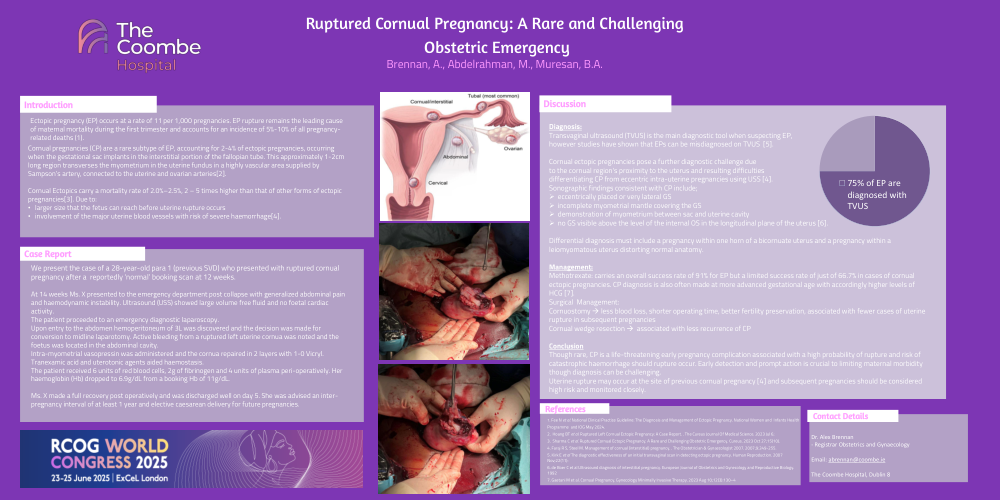
We present the case of a 28-year-old para 1 (previous SVD) who presented with ruptured cornual pregnancy after a reportedly ‘normal’ booking scan at 12 weeks.
At 14 weeks Ms. X presented to the emergency department post collapse with generalized abdominal pain and haemodynamic instability. Ultrasound (USS) showed large volume free fluid and no foetal cardiac activity.
The patient proceeded to an emergency diagnostic laparoscopy.
Upon entry to the abdomen hemoperitoneum of 3L was discovered and the decision was made for conversion to midline laparotomy. Active bleeding from a ruptured left uterine cornua was noted and the foetus was located in the abdominal cavity.
Intra-myometrial vasopressin was administered and the cornua repaired in 2 layers with 1-0 Vicryl. Tranexamic acid and uterotonic agents aided haemostasis.
The patient received 6 units of red blood cells, 2g of fibrinogen and 4 units of plasma peri-operatively. Her haemoglobin (Hb) dropped to 6.9g/dL from a booking Hb of 11g/dL.
Ms. X made a full recovery post operatively and was discharged well on day 5. She was advised an inter-pregnancy interval of at least 1 year and elective caesarean delivery for future pregnancies
Discussion:
Diagnosis:
Transvaginal ultrasound (TVUS) is the main diagnostic tool when suspecting EP,
however studies have shown that EPs can be misdiagnosed on TVUS [5].
Cornual ectopic pregnancies pose a further diagnostic challenge due
to the cornual region's proximity to the uterus and resulting difficulties
differentiating CP from eccentric intra-uterine pregnancies using USS [4].
Sonographic findings consistent with CP include;
Differential diagnosis must include a pregnancy within one horn of a bicornuate uterus and a pregnancy within a leiomyomatous uterus distorting normal anatomy.
Management:
Methotrexate: carries an overall success rate of 91% for EP but a limited success rate of just of 66.7% in cases of cornual ectopic pregnancies. CP diagnosis is also often made at more advanced gestational age with accordingly higher levels of HCG [7].
Surgical Management:
Cornuostomy à less blood loss, shorter operating time, better fertility preservation, associated with fewer cases of uterine rupture in subsequent pregnancies
Cornual wedge resection à associated with less recurrence of CP
Conclusion
Though rare, CP is a life-threatening early pregnancy complication associated with a high probability of rupture and risk of catastrophic haemorrhage should rupture occur. Early detection and prompt action is crucial to limiting maternal morbidity though diagnosis can be challenging.
Uterine rupture may occur at the site of previous cornual pregnancy [4] and subsequent pregnancies should be considered high risk and monitored closely.
Contact details:
Dr. Alex Brennan
- Registrar Obstetrics and Gynaecology
Email: abrennan@coombe.ie
The Coombe Hospital, Dublin 8