2,281 posters, 63 videos, 56 audios, 43 topics, 18 sessions, 6,174 authors, 2,323 institutions
ePostersLive by SciGen Technologies S.A. All rights reserved.
RCOG World Congress 2025
June 23 - 25, 2025 | ExCeL London, UK
EP.0191
Ipsilateral tubal stump ectopic pregnancy after partial salpingectomy: A case report
Poster Presenter
Authors
Affiliations
Part of Topic
Early pregnancy complications/early pregnancy and acute gynaecology
Title: Ipsilateral tubal stump ectopic pregnancy after partial salpingectomy: A case report
Objective: To highlight the challenges and learning in diagnosing and treating tubal stump ectopic pregnancy (EP) in high-risk patient.
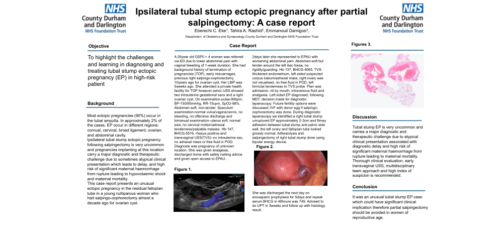
Case report: A 26year old G5P0 + 4 woman was referred via ED due to lower abdominal pain with vaginal bleeding of 1-week duration. She had background history of termination of pregnancies (TOP), early miscarriages, previous right salpingo-oophorectomy 10years ago for ovarian cyst. Her LMP was 5weeks ago. She attended a private health facility for TOP however pelvic USS showed two intrauterine gestational sacs and a right ovarian cyst. On examination-pulse-90bpm, BP-150/85mmhg, RR-15cpm, SpO2-98%. Abdomen-soft, non-tender. Speculum examination-normal vulva/vagina/cervix, no bleeding, no offensive discharge and bimanual examination–uterus soft, normal size, no cervical motion/adnexal tenderness/palpable masses. Hb-147, BHCG-5519, rhesus positive and transvaginal USS(TVS)–no intrauterine sac, no adnexal mass or free fluid in POD. Diagnosis was pregnancy of unknown location. She was given analgesia, discharged home with safety netting advice and given open access to EPAU.
2days later she represented to EPAU with worsening abdominal pain. Abdomen-soft but tender around the left iliac fossa, no rigidity/guarding. Hb-137, BHCG-4065, TVS-thickened endometrium, left sided suspected corpus luteum/adnexal mass, right ovary was not visualised, no free fluid in POD, left fornicial tenderness to TVS probe. Plan was admission, nil by mouth, intravenous fluid and analgesia. Left sided EP diagnosed, following MDT, decision made for diagnostic laparoscopy. Future fertility options were discussed; IVF with donor egg if salpingo-oophorectomy was done. During diagnostic laparoscopy we identified a right tubal stump unruptured EP approximately 2-3cm and flimsy adhesion between tubal stump and pelvic side wall, the left ovary and fallopian tube looked grossly normal. Adhesiolysis and salpingectomy of right tubal stump done using bipolar energy device. She was discharged the next day on enoxaparin prophylaxis for 5days and repeat serum BHCG in 48hours was 749. Advised to do UPT in 3weeks and follow up with histology result.
Discussion: Tubal stump EP is very uncommon and carries a major diagnostic and therapeutic challenge due to atypical clinical presentation associated with diagnostic delay and high risk of significant maternal haemorrhage from rupture leading to maternal mortality. Thorough clinical evaluation, early transvaginal USS, multidisciplinary team approach and high index of suspicion is recommended.
Conclusion: It was an unusual tubal stump EP case which could have significant clinical implication therefore partial salpingectomy should be avoided in women of reproductive age.