2,281 posters, 63 videos, 56 audios, 43 topics, 18 sessions, 6,174 authors, 2,323 institutions
ePostersLive by SciGen Technologies S.A. All rights reserved.
RCOG World Congress 2025
June 23 - 25, 2025 | ExCeL London, UK
EP.0153
Diagnostic Dilemmas of Uterine Arteriovenous Malformations (AVM)
Poster Presenter
Part of Topic
Early pregnancy complications/early pregnancy and acute gynaecology
Southend University
Hospital
NHS
NHS Foundation Trust
DIAGNOSTIC DILEMMAS OF UTERINE ARTERIOVENOUS MALFORMATIONS (AVM)
Author & Affiliations: RAISHAM SALEEM '(Clinical Fellow Obstetrics and Gynaecology)/SAROJINI BURUGAPALLI (Consultant Obstetrics and Gynaecology)
OBJECTIVES
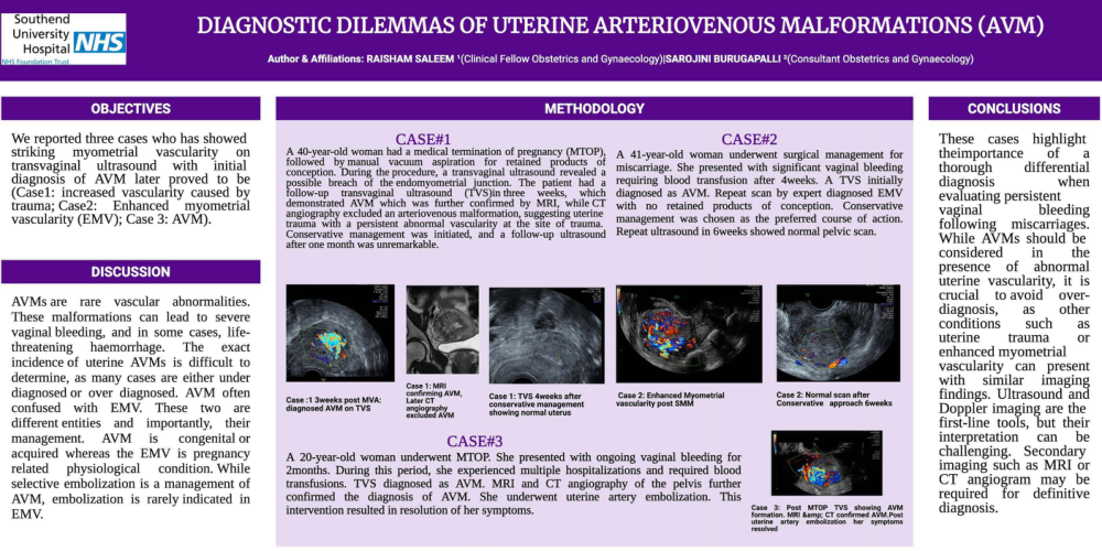
We reported three cases who has showed striking
myometrial
vascularity
on
transvaginal
ultrasound
with initial
diagnosis of AVM later proved to be (Casel: increased vascularity caused by trauma; Case2:
Enhanced
myometrial
vascularity (EMV); Case 3: AVM).
METHODOLOGY
CASE#1
CASE#2
followed by manual
A 40-year-old woman had a medical termination of pregnancy (MTOP):
A 41-year-old woman underwent surgical management for
vacuum aspiration for retained products
conception. During the procedure, a transvaginal ultrasound
miscarriage. She presented with significant vaginal bleeding
poisible breachs vas the endomsomet ial justin. The patien hahich requiring blood transfusion after 4weeks. A TVS initially
(TVS)in three
diagnosed as AVM. Repeat scan by expert diagnosed EMV
demonstrated AVM which was further confirmed by MRI, while CT angiography excluded an arteriovenous malformation, suggesting uterine
with no retained products of conception. Conservative
trauma with a persistent abnormal vascularity at the site of trauma.
management was chosen as the preferred course of action.
Conservative management was initiated, and a follow-up ultrasound Repeat ultrasound in weeks showed normal pelvic scan. after one month was unremarkable.
DISCUSSION
AVMs are
rare
vascular abnormalities.
These malformations can lead to severe vaginal bleeding, and in some cases, life-threatening haemorrhage.
The exact
incidence of uterine AVMs is difficult to determine, as many cases are either under diagnosed or over diagnosed. AVM often confused with EMV. These two are
different entities and importantly, their management.
AVM is
congenital or
acquired whereas the EMV is pregnancy related physiological
condition. While
selective embolization is a management of
AVM, embolization is rarely indicated in EMV.
Case :1 3weeks post MVA: diagnosed AVM on TVS
Case 1: MRI
confirming AVM,
Later CT
angiography excluded AVM
Case 1: TVS 4weeks after conservative management showing normal uterus
CASE#3
A 20-year-old woman underwent MTOP. She presented with ongoing vaginal bleeding for 2months. During this period, she experienced multiple hospitalizations and required blood transfusions. TVS diagnosed as AVM. MRI and CT angiography of the pelvis further confirmed the diagnosis of AVM. She underwent uterine artery embolization. This
intervention resulted in resolution of her symptoms.
Case 2: Enhanced Myometrial vascularity post SMM
Case 2: Normal scan after Conservative
approach 6weeks
CONCLUSIONS
These cases highlight theimportance
of
thorough differential diagnosis
when
evaluating persistent vaginal
bleeding
following miscarriages.
While AVMs should be considered
in the
presence
of abnormal
uterine vascularity, it is crucial to avoid over-
diagnosis,
as
other
conditions
such
as
uterine
trauma
or
enhanced myometrial vascularity can present with similar imaging findings. Ultrasound and Doppler imaging are the first-line tools, but their interpretation can be
challenging.
Secondary
imaging such as MRI or angiogram may be
required
for definitive
diagnosis.
Case 3:
Post
MTOP
TVS showing
AVM
formation. MRI &: CT confirmed AVM.Post uterine artery embolization
symptoms