2,281 posters, 63 videos, 56 audios, 43 topics, 18 sessions, 6,174 authors, 2,323 institutions
ePostersLive by SciGen Technologies S.A. All rights reserved.
RCOG World Congress 2025
June 23 - 25, 2025 | ExCeL London, UK
EP.1303
Cardiovascular arrest during caesarean section: a case of venous air embolism
Poster Presenter
Authors
Hong Jing Lee, Santi Wei Che Lim, Aaron Yik Hang Kong, Chik-Foo Yim, Ann Wright
Affiliations
Part of Topic
Imaging in obstetrics
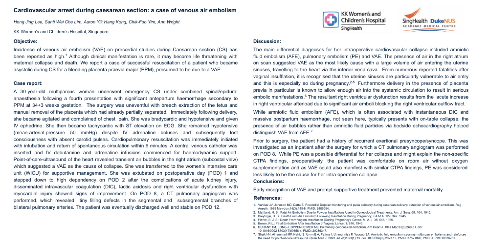
Cardiovascular arrest during caesarean section: a case of venous air embolism
Hong Jing Lee, Santi Wei Che Lim, Aaron Yik Hang Kong, Chik-Foo Yim, Ann Wright
KK Women’s and Children’s Hospital, Singapore
Objective:
Incidence of venous air embolism (VAE) on precordial studies during Caesarean section (CS) has been reported as high.1 Although clinical manifestation is rare, it may become life threatening with maternal collapse and death. We report a case of successful resuscitation of a patient who became asystolic during CS for a bleeding placenta praevia major (PPM), presumed to be due to a VAE.
Case report:
A 30-year-old multiparous woman underwent emergency CS under combined spinal/epidural anaesthesia following a fourth presentation with significant antepartum haemorrhage secondary to PPM at 34+3 weeks gestation. The surgery was uneventful with breech extraction of the fetus and manual removal of the placenta which had already partially separated. Immediately following delivery, she became agitated and complained of chest pain. She was bradycardic and hypotensive and given IV ephedrine. She then became tachycardic with ST elevation on ECG. She remained hypotensive (mean-arterial-pressure 50 mmHg) despite IV adrenaline boluses and subsequently lost consciousness with absent carotid pulses. Cardiopulmonary resuscitation was immediately initiated with intubation and return of spontaneous circulation within 6 minutes. A central venous catheter was inserted and IV dobutamine and adrenaline infusions commenced for haemodynamic support. Point-of-care-ultrasound of the heart revealed transient air bubbles in the right atrium (subcostal view) which suggested a VAE as the cause of collapse. She was transferred to the women’s intensive care unit (WICU) for supportive management. She was extubated on postoperative day (POD) 1 and stepped down to high dependency on POD 2 after the complications of acute kidney injury, disseminated intravascular coagulation (DIC), lactic acidosis and right ventricular dysfunction with myocardial injury showed signs of improvement. On POD 8, a CT pulmonary angiogram was performed, which revealed tiny filling defects in the segmental and subsegmental branches of bilateral pulmonary arteries. The patient was eventually discharged well and stable on POD 12.
Discussion:
The main differential diagnoses for her intraoperative cardiovascular collapse included amniotic fluid embolism (AFE), pulmonary embolism (PE) and VAE. The presence of air in the right atrium on scan suggested VAE as the most likely cause with a large volume of air entering the uterine sinuses, travelling to the heart via the inferior vena cava. From numerous reported fatalities after vaginal insufflation, it is recognised that the uterine sinuses are particularly vulnerable to air entry and this is especially so during pregnancy.2-5 Furthermore delivery in the presence of placenta previa in particular is known to allow enough air into the systemic circulation to result in serious embolic manifestations.6 The resultant right ventricular dysfunction results from the acute increase in right ventricular afterload due to significant air emboli blocking the right ventricular outflow tract.
While amniotic fluid embolism (AFE), which is often associated with instantaneous DIC and massive postpartum haemorrhage, not seen here, typically presents with on-table collapse, the presence of air bubbles rather than amniotic fluid particles via bedside echocardiography helped distinguish VAE from AFE.7
Prior to surgery, the patient had a history of recurrent exertional presyncope/syncope. This was investigated as an inpatient after the surgery for which a CT pulmonary angiogram was performed on POD 8. While PE was a possible differential for her collapse and might explain the non-specific CTPA findings, preoperatively, the patient was comfortable on room air without oxygen supplementation and as VAE could also manifest with similar CTPA findings, PE was considered less likely to be the cause for her intra-operative collapse.
Conclusions:
Early recognition of VAE and prompt supportive treatment prevented maternal mortality.
References:
-
Vartikar JV, Johnson MD, Datta S. Precordial Doppler monitoring and pulse oximetry during cesarean delivery: detection of venous air embolism. Reg Anesth. 1989 May-Jun;14(3):145-8. PMID: 2486594.
-
Martland, H. S.: Fatal Air Embolism Due to Powder Insufflations Used in Gynecological Treatments, Am. J. Surg. 68: 164, 1945.
-
Breyfogle, H. S.: Death From Air Embolism Following Insufflation During Pregnancy, J.A.M.A. 129: 342. 1945.
-
Pierce, S. J. S.: Death From Vaginal Insufflation (During Pregnancy), Canad. M. A. J. 35: 668, 1936.
-
Brown, R.L.: Fatal Embolism After Insufflation of Vagina, Lancet 1: 616, 1943.
-
DURANT TM, LONG J, OPPENHEIMER MJ. Pulmonary (venous) air embolism. Am Heart J. 1947 Mar;33(3):269-81. doi: 10.1016/0002-8703(47)90656-x. PMID: 20286347.
-
Shaikh N, Alhammad MF, Nahid S, Umm E A, Fatima I, Ummunnisa F, Yaqoub SA. Amniotic fluid embolism causing multiorgan embolisms and reinforces the need for point-of-care ultrasound. Qatar Med J. 2023 Jul 28;2023(1):13. doi: 10.5339/qmj.2023.13. PMID: 37521090; PMCID: PMC10376761.