2,281 posters, 63 videos, 56 audios, 43 topics, 18 sessions, 6,174 authors, 2,323 institutions
ePostersLive by SciGen Technologies S.A. All rights reserved.
RCOG World Congress 2025
June 23 - 25, 2025 | ExCeL London, UK
EP.1299
Artificial Intelligence Identification of Key Sonographic Landmarks in the Fetal Brain
Poster Presenter
Authors
Abhiram Kanneganti, Jemima Teo, Yihao Ai, Rachel Ern Si Goh, Xingyi Yang, Cece Dimech, Arundhati Gosavi, Biswas Arijit
Affiliations
Department of Obstetrics and Gynaecology, National University of Hospital, Department of Electrical and Computer Engineering, College of Design and Engineering, National University of Singapore, Yong Loo Lin School of Medicine, National University of Singapore
Part of Topic
Imaging in obstetrics
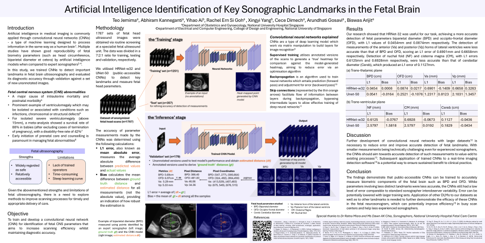
Introduction: Artificial intelligence in medical imaging is commonly applied through convolutional neural networks (CNNs) - a type of machine learning designed to process information in the same way as a human brain. Multiple studies have shown good reproducibility of fetal biometry parameters (such as head circumference, biparietal diameter diameter et cetera) by artificial intelligence models when compared to expert sonographers. In this study, we trained CNNs to detect important landmarks in fetal brain ultrasonography and evaluated its diagnostic accuracy through validation against a set of new unannotated images. Fetal central nervous system (CNS) abnormalities are a major cause of intrauterine mortality and postnatal morbidity. Prominent example of ventriculomegaly which may be isolated or associated with conditions such as infections, chromosomal or structural defects. For isolated severe ventriculomegaly (above 15mm), a meta-analysis showed a survival rate of 88% in babies (after excluding cases of termination of pregnancy), with a disability-free rate of 42%. Early initiation of prenatal care and counselling is paramount in managing fetal abnormalities.
Objective: To train and develop a convolutional neural network (CNN) for identification of fetal CNS parameters that aims to increase scanning efficiency whilst maintaining diagnostic accuracy.
Methodology: 1787 sets of fetal head ultrasound images were obtained via routine screening at a specialist fetal ultrasound unit. The data was divided in a 7:2:1 ratio for training, testing and validation, respectively. We utilised HRNet-w32 and UNet-50 (public-accessible CNNs) to detect key landmarks and measure fetal head parameters. The accuracy of parameter measurements made by the CNNs was determined using the following calculations: L1 error, also known as mean absolute error, measures the average absolute difference between predicted values and actual values. Bias calculates the mean difference between ground truth distance and estimated distance for all measurements (not the absolute value), providing an indication of how robust the estimation is.
Results: Our research showed that HRNet-32 was useful for our task, achieving a more accurate detection of fetal parameters biparietal diameter (BPD) and occipito-frontal diameter (OFD), with L1 values of 0.0454mm and 0.0874mm respectively. The detection of measurements of the anterior (Va) and posterior (Vp) horns of lateral ventricles were less accurate than that of BPD and OFD, scoring an L1 error of 0.6901mm and 0.6658mm respectively. Detection of nuchal fold (NF) and cisterna magna (CM), with L1 errors 0.6125mm and 0.6928mm respectively, were less accurate than that of cerebellar diameter (Cereb), which produced an L1 error of 0.1127mm.
Discussion: Further development of convolutional neural networks with larger datasets is necessary to reduce error and improve accurate detection of fetal landmarks. With smaller measurements being technically challenging even for experienced sonographers, the CNNs should aim towards accurate detection of such measurements to value-add to existing processes. Subsequent application of trained CNNs to a real-time imaging detection software is a potential way to ensure sustained benefit to clinical practice.
Conclusion: The findings demonstrate that public-accessible CNNs can be trained to accurately measure biometric components of the fetal brain such as BPD and OFD. While parameters involving less distinct landmarks were less accurate, the CNNs still had a low level of error comparable to standard sonographer interobserver variability. Error can be potentially lowered with larger training sets. Application of other DLMs to our datasets as centers and help less experienced sonographers.