2,281 posters, 63 videos, 56 audios, 43 topics, 18 sessions, 6,174 authors, 2,323 institutions
ePostersLive by SciGen Technologies S.A. All rights reserved.
RCOG World Congress 2025
June 23 - 25, 2025 | ExCeL London, UK
EP.1296
Fetal MRI estimates fetal weight centiles closer to birthweight than ultrasound estimates
Poster Presenter
Part of Topic
Imaging in obstetrics
Fetal MRI Estimates Fetal Weight Centiles Closer To True Birthweight Than Ultrasound
1. Background
Correctly estimating fetal size= Clinically important
Antenatal fetal size = Estimated Fetal Weight (EFW)
Current gold standard is Ultrasound Scan (US)
Both Small and Large for Gestational Age (SGA, LGA,) fetus
→ Birth complications
As per Big Baby Trial, diagnosis of LGA by US has large variation
& false positives 60% (Gardosi et al., 2025)
Scope for improving diagnostic accuracy, particularly in the LGA
cohort.
There is emerging evidence that MRI may be more accurate
at predicting fetal weight compared to US.
2.1. Methods
As part of the currently recruiting MibirthResearch Study
(www.mibirthstudy.com) 131 singleton participants,
between 35+5 and 40+6 weeks gestation,
had the following within 48 hours of each other:
1. Antenatal fetal growth USS
2. Low field MRI (0.55Tesla)
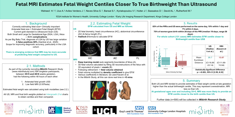
Estimated fetal weight was calculated using both modalities (see 2.2.)
All US, MRI and final birth weights plotted on Intergrowth 21st charts
to obtain centiles and then compared.
5. Summary
Both US and MRI tended to provide estimates of fetal weight centile at late gestation
higher than the actual birthweight centile. This may represent overestimation, MRI
less so than US.
At gestational ages over and including 35+5, MRI was more likely to provide an
EFW centile closer to the birth centile than US.
Further data (n=500) will be collected in Mibirth Research Study.