2,281 posters, 63 videos, 56 audios, 43 topics, 18 sessions, 6,174 authors, 2,323 institutions
ePostersLive by SciGen Technologies S.A. All rights reserved.
RCOG World Congress 2025
June 23 - 25, 2025 | ExCeL London, UK
EP.1288
Abdominal-pelvic abscess postpartum, a rare event, occurred in 2022-2024 at Nizwa hospital.
Poster Presenter
Part of Topic
Hot topics (emerging research/issues) in obstetrics
Abdominal-pelvic abscess postpartum, a rare event, occurred in 2022-2024 at Nizwa hospital. Rekha B Venkatesh, Shadya Al Saeghi, Hansa Dhar, Uma U Singh,Sultan Ali Al Busaidi, Qamariya Khalfan Saud Ambusaidi Department of Obstetrics and Gynaecology, Nizwa Hospital, Oman. Department of Radiology, Nizwa Hospital, Oman.
Introduction-- Intraabdominal infection (IAI) is a pocket of pus or infected fluid located within the abdominal cavity. IAI “particularly if not well managed” are recognized as major contributors to mortality in emergency departments globally 1.Infections can be within the pelvis, around the operation site, or in the organs. These infections lead to serious condition, increase maternal morbidity, lengthen hospital stay, increase medical costs and can increase maternal mortality tremendously. (32.5%)2. Subsequently requring a complicated course of management in high dependancy/intensive care units, ultrasound guided drainage of pus or sometimes surgical re-exploration or even hysterectomy. We report a few cases of abscess in our set up which required various course of management.
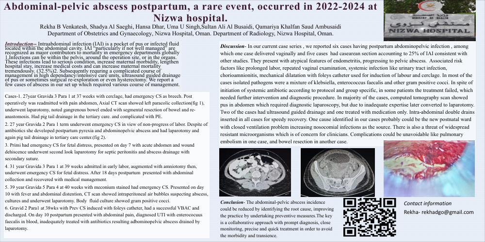
Cases-1. 27year Gravida 3 Para 1 at 37 weeks with cerclage, had emergency CS as breech. Post operatively was readmitted with pain abdomen, Axial CT scan showed left paracolic collection(fig 1), underwent laparotomy, noted gangrenous bowel ended with segmental resection of bowel and re-anastomosis. Had pig tail drainage in the tertiary care. and complicated with PE. 2. 27 year Gravida 2 Para 1 term underwent emergency CS in view of non-progress of labor. Despite of antibiotics she developed postpartum pyrexia and abdominopelvic abscess and had laparotomy and again pig tail drainage in tertiary care center.(fig 2). 3. Primi had emergency CS for fetal distress, presented on day 7 with acute abdomen and wound dehiscence underwent second look laparotomy for septic peritonitis and abscess drainage with secondary suture. 4. 31 year Gravida 3 Para 1 at 39 weeks admitted in early labor, augmented with amniotomy then, underwent emergency CS for fetal distress. After 18 days postpartum presented with abdominal collection and recovered with medical management. 5. 39 year Gravida 5 Para 4 at 40 weeks with meconium stained had emergency CS. Presented on day 10 with fever and abdominal distention, CT scan showed intraperitoneal air bubbles suspecting abscess, cultures and underwent laparotomy. Body fluid culture showed gram positive cocci. 6. Gravid 2 Para1 at 38wks with Prev CS induced with foleys catheter, had a successful VBAC and discharged. On day 10 postpartum presented with abdominal pain, diagnosed UTI with enterococuus faecalis in blood, inadequately treated with antibiotics resulting adbominopelvic abscess drained by laparotomy.
Discussion- In our current case series , we reported six cases having postpartum abdominopelvic infection , among which one case delivered vaginally and five cases had casearean section accounting to 25% of IAI consistent with other studies. They present with atypical features of endometritis, progressing to pelvic abscess. Associated risk factors like prolonged labor, repeated vaginal examination, systemic infection like urinary tract infection, chorioamnionitis, mechanical dilatation with foleys catheter used for induction of labour and cerclage. In most of the cases isolated pathogens were a mixture of klebsiella, enterococcus faecalis and other gram positive cocci. In spite of initiation of systemic antibiotic according to protocol and group specific, in some patients the treatment failed, which needed further intervention and diagnostic procedure. In majority of the cases, computed tomography scan showed pus in abdomen which required diagnostic laparoscopy, but due to inadequate expertise later converted to laparotomy. Two of the cases had ultrasound guided drainage and one treated with medication only. Intra-abdominal double drains inserted in all cases for speedy recovery. One cause identified in our cases probably could be the new postnatal ward with closed ventilation problem increasing nosocomial infections as the source. There is also a threat of widespread resistant microorganisms which is of concern for clinicians. Complications could be unavoidable like pulmonary embolism in one case, and bowel resection in another case.
Conclusion- The abdominal-pelvic abscess incidence could be reduced by identifying the root cause, improving the practice by undertaking preventive measures.The key is a collaborative approach with prompt diagnosis, close monitoring, precise and quick treatment in order to avoid the morbidity and transience.