2,281 posters, 63 videos, 56 audios, 43 topics, 18 sessions, 6,174 authors, 2,323 institutions
ePostersLive by SciGen Technologies S.A. All rights reserved.
RCOG World Congress 2025
June 23 - 25, 2025 | ExCeL London, UK
EP.1283
Case Report: A very rare case of post-partum Adrenal Haematoma
Poster Presenter
Authors
Affiliations
Part of Topic
Hot topics (emerging research/issues) in obstetrics
CASE REPORT:
A Very Rare Case of Postpartum Adrenal Haematoma
Dr Priya Abraham1 ST2 in Obstetrics & Gynaecology, Priya.Abraham1@nhs.net
Miss Farah Naz1 Consultant Obstetrician & Gynaecologist,
Mr Hashem Elhossamey1 Consultant Obstetrician & Gynaecologist
1. The Dudley Group NHS Foundation Trust
OBJECTIVES
BACKGROUND
Adrenal haemorrhage (AH) is a rare condition, with an incidence of 0.14–1.8% during pregnancy, and even fewer cases reported in the puerperium[1,2]. It often presents with non-specific signs, which can delay diagnosis and lead to life-threatening complications such as adrenal crisis, shock, and, if undiagnosed, potentially death[1,2].
The aetiology remains unclear but is believed to be associated with adrenocorticotropic hormone (ACTH), adrenal vein thrombosis, postpartum haemorrhage, anti-phospholipid syndrome, and the increased hypercoagulability of pregnancy[1,3].
Incidence may be higher in the presence of pregnancy-related adrenal hyperplasia and limited venous drainage. In cases of retroperitoneal haematomas following delivery, AH should be considered as a differential diagnosis[3,4].Outside of pregnancy, the primary causes of AH include trauma and adjacent tumours[2].
Damage to the adrenal glands can result in deficiencies in aldosterone, cortisol, androgens, and catecholamines—all of which are essential for maintaining homeostasis and regulating maternal and foetal haemodynamics[1].The most common presentation is non-specific pain in the epigastric, flank, upper or lower back, or pelvic regions, occurring in 65–85% of cases[1,2].
Symptoms of adrenal insufficiency may also be present, such as fatigue, weakness, dizziness, and anorexia[1]. In severe cases, this may progress to a life-threatening adrenal crisis or Addison’s disease. If left untreated, patients may develop hypotension, tachycardia, shock, and, in extreme cases, maternal and foetal death[1]. Imaging is essential for diagnosis; however, blood tests may also reveal hyponatraemia and hypoglycaemia[3].
CASE PRESENTATION
A 25-year-old patient with a history of three miscarriages and two live births. The patient underwent category 1 caesarean section for foetal distress and labour dystocia at 38+1 weeks gestation. The patient had maternal risk factors including a Body Mass Index (BMI) of 48, positive for Group B streptococcal infection, previous venous thrombotic event requiring antenatal low molecular weight heparin (LMWH), previous pre-eclampsia requiring antenatal aspirin and previous gestational diabetes.
Postoperatively the patient developed a wound infection and haematoma. She was managed medically with antibiotics including flucloxacillin and metronidazole. The patient also received input from tissue viability for wound care and was discharged once deemed well.
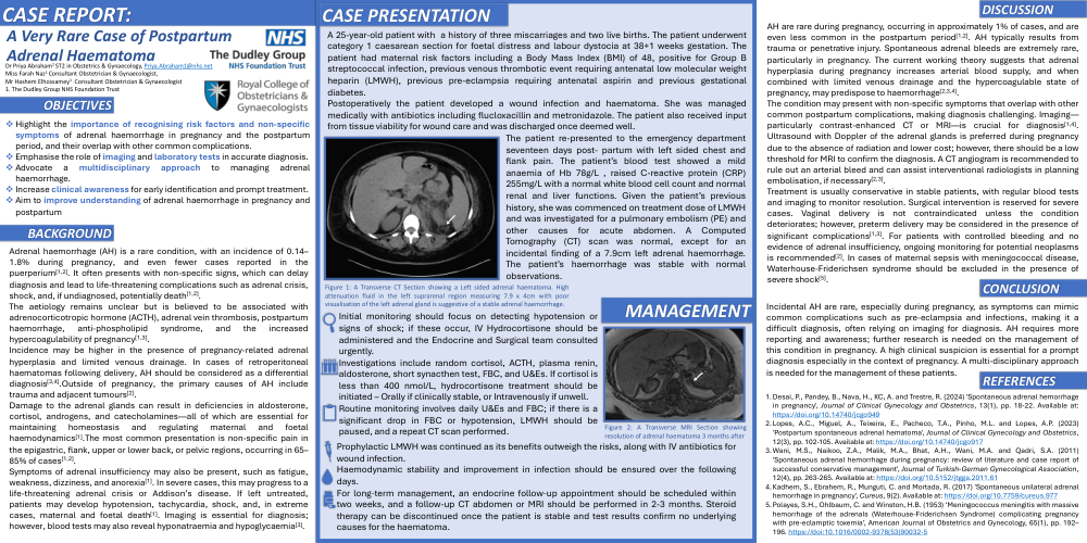
The patient re-presented to the emergency department seventeen days post- partum with left sided chest and flank pain. The patient’s blood test showed a mild anaemia of Hb 78g/L , raised C-reactive protein (CRP) 255mg/L with a normal white blood cell count and normal renal and liver functions. Given the patient’s previous history, she was commenced on treatment dose of LMWH and was investigated for a pulmonary embolism (PE) and other causes for acute abdomen. A Computed Tomography (CT) scan was normal, except for an incidental finding of a 7.9cm left adrenal haemorrhage. The patient’s haemorrhage was stable with normal observations.
Figure 1: A Transverse CT Section showing a Left sided adrenal haematoma. High attenuation fluid in the left suprarenal region measuring 7.9 x 4cm with poor visualisation of the left adrenal gland is suggestive of a stable adrenal haemorrhage.
Figure 2: A Transverse MRI Section showing resolution of adrenal haematoma 3 months after
MANAGEMENT
DISCUSSION
AH are rare during pregnancy, occurring in approximately 1% of cases, and are even less common in the postpartum period[1,2]. AH typically results from trauma or penetrative injury. Spontaneous adrenal bleeds are extremely rare, particularly in pregnancy. The current working theory suggests that adrenal hyperplasia during pregnancy increases arterial blood supply, and when combined with limited venous drainage and the hypercoagulable state of pregnancy, may predispose to haemorrhage[2,3,4].
The condition may present with non-specific symptoms that overlap with other common postpartum complications, making diagnosis challenging. Imaging—particularly contrast-enhanced CT or MRI—is crucial for diagnosis[1,4]. Ultrasound with Doppler of the adrenal glands is preferred during pregnancy due to the absence of radiation and lower cost; however, there should be a low threshold for MRI to confirm the diagnosis. A CT angiogram is recommended to rule out an arterial bleed and can assist interventional radiologists in planning embolisation, if necessary[2,3].
Treatment is usually conservative in stable patients, with regular blood tests and imaging to monitor resolution. Surgical intervention is reserved for severe cases. Vaginal delivery is not contraindicated unless the condition deteriorates; however, preterm delivery may be considered in the presence of significant complications[1,3]. For patients with controlled bleeding and no evidence of adrenal insufficiency, ongoing monitoring for potential neoplasms is recommended[2]. In cases of maternal sepsis with meningococcal disease, Waterhouse-Friderichsen syndrome should be excluded in the presence of severe shock[5].
CONCLUSION
Incidental AH are rare, especially during pregnancy, as symptoms can mimic common complications such as pre-eclampsia and infections, making it a difficult diagnosis, often relying on imaging for diagnosis. AH requires more reporting and awareness; further research is needed on the management of this condition in pregnancy. A high clinical suspicion is essential for a prompt diagnosis especially in the context of pregnancy. A multi-disciplinary approach is needed for the management of these patients.
REFERENCES