2,281 posters, 63 videos, 56 audios, 43 topics, 18 sessions, 6,174 authors, 2,323 institutions
ePostersLive by SciGen Technologies S.A. All rights reserved.
RCOG World Congress 2025
June 23 - 25, 2025 | ExCeL London, UK
EP.1117
A Case Report: Giant Placental Chorioangioma with Favourable Outcome
Poster Presenter
Part of Topic
High risk pregnancy
A Case Report:Giant Placental Chorioangioma with Favourable Outcome
Authors: Miss Shamsa Kassam, Dr Rabia Haq, Mr Shyam Das
Institution: Luton and Dunstable Foundation Trust, Bedfordshire Hospitals
Introduction Chorioangioma is the most common benign tumour of the placenta, characterized by arteriovenous (AV) shunting. While small chorioangiomas (<1% incidence) are generally asymptomatic, larger lesions (>4 cm) are rare and associated with significant pregnancy complications, including polyhydramnios, fetal hydrops, cardiomegaly, fetal anaemia, congestive heart failure, intrauterine death, antepartum haemorrhage, and preterm delivery. This case report highlights a rare instance of a giant placental chorioangioma with a favourable outcome.
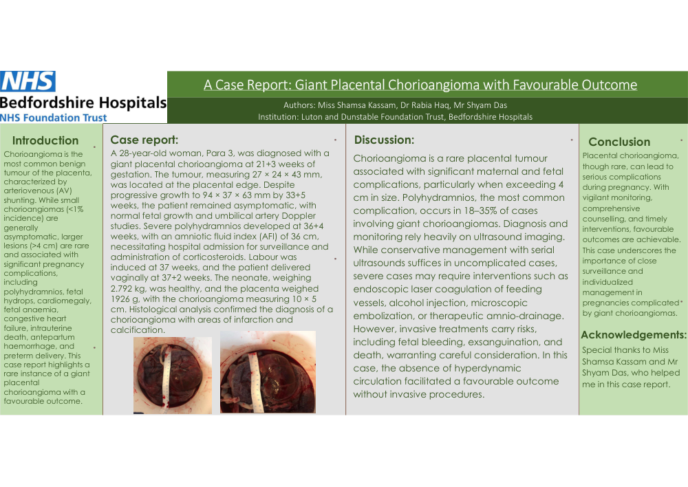
Case report: A 28-year-old woman, Para 3, was diagnosed with a giant placental chorioangioma at 21+3 weeks of gestation. The tumour, measuring 27 × 24 × 43 mm, was located at the placental edge. Despite progressive growth to 94 × 37 × 63 mm by 33+5 weeks, the patient remained asymptomatic, with normal fetal growth and umbilical artery Doppler studies. Severe polyhydramnios developed at 36+4 weeks, with an amniotic fluid index (AFI) of 36 cm, necessitating hospital admission for surveillance and administration of corticosteroids. Labour was induced at 37 weeks, and the patient delivered vaginally at 37+2 weeks. The neonate, weighing 2.792 kg, was healthy, and the placenta weighed 1926 g, with the chorioangioma measuring 10 × 5 cm. Histological analysis confirmed the diagnosis of a chorioangioma with areas of infarction and calcification.
Discussion: Chorioangioma is a rare placental tumour associated with significant maternal and fetal complications, particularly when exceeding 4 cm in size. Polyhydramnios, the most common complication, occurs in 18–35% of cases involving giant chorioangiomas. Diagnosis and monitoring rely heavily on ultrasound imaging. While conservative management with serial ultrasounds suffices in uncomplicated cases, severe cases may require interventions such as endoscopic laser coagulation of feeding vessels, alcohol injection, microscopic embolization, or therapeutic amnio-drainage. However, invasive treatments carry risks, including fetal bleeding, exsanguination, and death, warranting careful consideration. In this case, the absence of hyperdynamic circulation facilitated a favourable outcome without invasive procedures.
Conclusion: Placental chorioangioma, though rare, can lead to serious complications during pregnancy. With vigilant monitoring, comprehensive counselling, and timely interventions, favourable outcomes are achievable. This case underscores the importance of close surveillance and individualized management in pregnancies complicated by giant chorioangiomas. Acknowledgements: Special thanks to Miss Shamsa Kassam and Mr Shyam Das, who helped me in this case report.