2,281 posters, 63 videos, 56 audios, 43 topics, 18 sessions, 6,174 authors, 2,323 institutions
ePostersLive by SciGen Technologies S.A. All rights reserved.
RCOG World Congress 2025
June 23 - 25, 2025 | ExCeL London, UK
EP.1100
Successful management of atypical presentation of Sub-capsular Liver Hematoma with peri-hepatic packing.
Poster Presenter
Authors
Affiliations
Royal Berkshire Hospital, Royal Derby Hospital, Princess Anne Hospital
Part of Topic
High risk pregnancy
Successful management of atypical presentation of Sub-capsular Liver Hematoma with peri-hepatic packing.
Objective: This case report describes the successful management of an atypical presentation of sub-capsular liver hematoma (SLH) with peri-hepatic packing in a district general hospital.
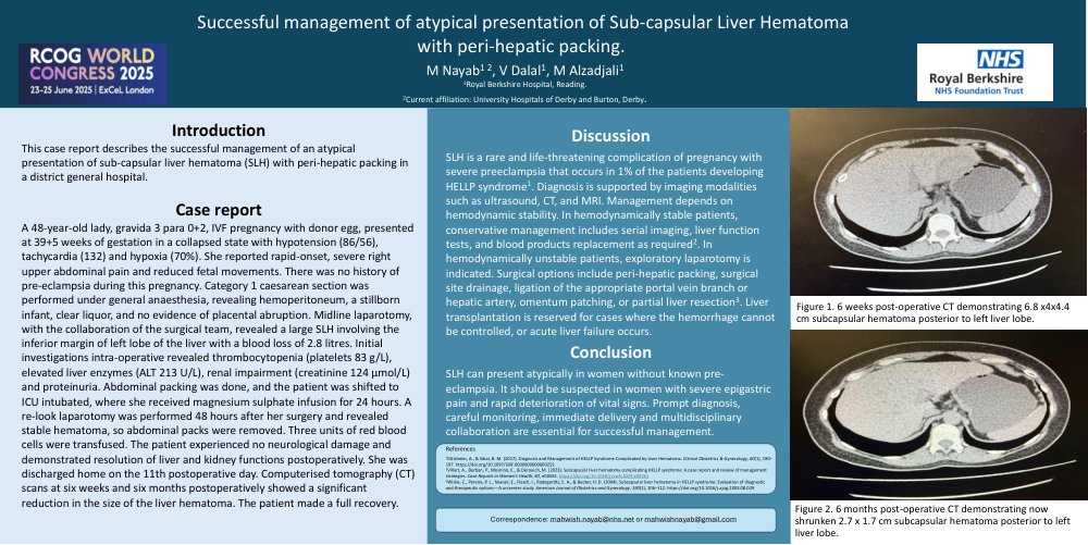
Case report: A 48-year-old lady, gravida 3 para 0+2, IVF pregnancy with donor egg, presented at 39+5 weeks of gestation in a collapsed state with hypotension (86/56), tachycardia (132) and hypoxia (70%). She reported rapid-onset, severe right upper abdominal pain and reduced fetal movements. There was no history of pre-eclampsia during this pregnancy. Category 1 caesarean section was performed under general anaesthesia, revealing hemoperitoneum, a stillborn infant, clear liquor, and no evidence of placental abruption. Midline laparotomy, with the collaboration of the surgical team, revealed a large SLH involving the inferior margin of left lobe of the liver with a blood loss of 2.8 litres. Initial investigations intra-operative revealed thrombocytopenia (platelets 83 g/L), elevated liver enzymes (ALT 213 U/L), renal impairment (creatinine 124 µmol/L) and proteinuria. Abdominal packing was done, and the patient was shifted to ICU intubated, where she received magnesium sulphate infusion for 24 hours. A re-look laparotomy was performed 48 hours after her surgery and revealed stable hematoma, so abdominal packs were removed. Three units of red blood cells were transfused. The patient experienced no neurological damage and demonstrated resolution of liver and kidney functions postoperatively. She was discharged home on the 11th postoperative day. Computerised tomography (CT) scans at six weeks and six months postoperatively showed a significant reduction in the size of the liver hematoma. The patient made a full recovery.
Discussion: SLH is a rare and life-threatening complication of pregnancy with severe preeclampsia that should ideally be managed in tertiary care centres. Diagnosis is supported by imaging modalities such as ultrasound, CT, and MRI. Management depends on hemodynamic stability. In hemodynamically stable patients, conservative management includes serial imaging, liver function tests, and blood products replacement as required. In hemodynamically unstable patients, exploratory laparotomy is indicated. Surgical options include peri-hepatic packing, surgical site drainage, ligation of the appropriate portal vein branch or hepatic artery, omentum patching, or partial liver resection. Liver transplantation is reserved for cases where the hemorrhage cannot be controlled, or acute liver failure occurs.
Conclusion: SLH can present atypically in women without known pre-eclampsia. It should be suspected in women with severe epigastric pain and rapid deterioration of vital signs. Prompt diagnosis, careful monitoring, immediate delivery and multidisciplinary collaboration are essential for successful management.