2,281 posters, 63 videos, 56 audios, 43 topics, 18 sessions, 6,174 authors, 2,323 institutions
ePostersLive by SciGen Technologies S.A. All rights reserved.
RCOG World Congress 2025
June 23 - 25, 2025 | ExCeL London, UK
EP.1098
Spontaneous liver rupture secondary to severe pre-eclampsia: a case report
Poster Presenter
Authors
Affiliations
Part of Topic
High risk pregnancy
Title:
Spontaneous liver rupture in pregnancy with complex medical and social history
Objectives:
Spontaneous liver rupture in pregnancy is a rare but grave complication with high maternal and fetal mortality. It is most often associated with severe preeclampsia, HELLP syndrome or acute fatty liver of pregnancy. We report a case that highlights the challenges of managing this acute life-threatening condition in a 34-week pregnant lady with complex social and past medical history.
Case summary:
36 years old , P1 (NVD 13 years ago), BMI of 35, late booker at 28 weeks (recent migrant). Non-English speaking. History of breast cancer managed with lumpectomy, chemotherapy and radiotherapy 3 years prior and advised to take 5 years of Tamoxifen. Unfortunately, due to her status she struggled to get her appropriate medication and fell pregnant.
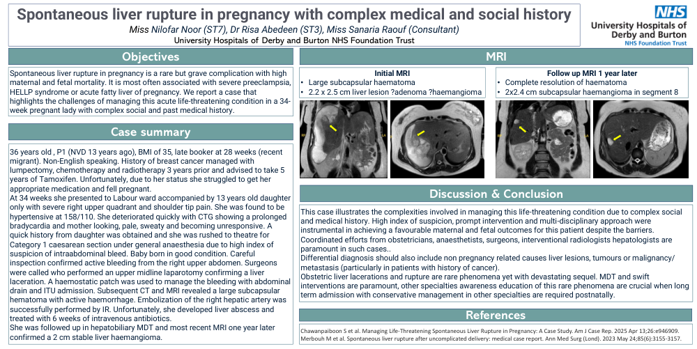
At 34 weeks she presented to Labour ward accompanied by 13 years old daughter only with severe right upper quadrant and shoulder tip pain. She was found to be hypertensive at 158/110. She deteriorated quickly withCTG showing a prolonged bradycardia and mother looking, pale, sweaty and becoming unresponsive. A quick history from daughter was obtained and she was rushed to theatre for Category 1 caesarean section under general anaesthesia due to high index of suspicion of intraabdominal bleed. Baby born in good condition. Careful inspection confirmed active bleeding from the right upper abdomen. Surgeons were called who performed an upper midline laparotomy confirming a liver laceration. A haemostatic patch was used to manage the bleeding with abdominal drain and ITU admission. Subsequent CT and MRI revealed a large subcapsular hematoma with active haemorrhage. Embolization of the right hepatic artery was successfully performed by IR. Unfortunately, she developed liver abscess and treated with 6 weeks of intravenous antibiotics.
She was followed up in hepatobiliary MDT and most recent MRI one year later confirmed a 2 cm stable liver haemangioma.
Discussion and Conclusion:
This case illustrates the complexities involved in managing this life-threatening condition due to complex social and medical history. High index of suspicion, prompt intervention and multi-disciplinary approach were instrumental in achieving a favourable maternal and fetaloutcomes for this patient despite the barriers.
Coordinated efforts from obstetricians, anaesthetists, surgeons, interventional radiologists hepatologists are paramount in such cases..
Differential diagnosis should also include non pregnancy related causes liver lesions, tumours or malignancy/ metastasis (particularly in patients with history of cancer).
Obstetric liver lacerations and rupture are rare phenomena yet with devastating sequel. MDT and swift interventions are paramount, other specialties awareness education of this rare phenomena are crucial when long term admission with conservative management in other specialties are required postnatally.
References:
Chawanpaiboon S et al. Managing Life-Threatening Spontaneous Liver Rupture in Pregnancy: A Case Study. Am J Case Rep. 2025 Apr 13;26:e946909.
Merbouh M et al. Spontaneous liver rupture after uncomplicated delivery: medical case report. Ann Med Surg(Lond). 2023 May 24;85(6):3155-3157.