2,281 posters, 63 videos, 56 audios, 43 topics, 18 sessions, 6,174 authors, 2,323 institutions
ePostersLive by SciGen Technologies S.A. All rights reserved.
RCOG World Congress 2025
June 23 - 25, 2025 | ExCeL London, UK
EP.1093
Missed Diagnosis of Vasa Praevia Leading to Postnatal Discovery: A Case Report
Poster Presenter
Authors
Anusuya Bansal, Zahra Anwar, Deniz Al-Hirmizy, Maja Kotlinska
Affiliations
Part of Topic
High risk pregnancy
Missed Diagnosis of Vasa Praevia Leading to Postnatal Discovery: A Case Report
Dr Anusuya Bansal, Dr Zahra Anwar, Ms Deniz Al-Hirmizy, Ms Maja Kotlinska.
Diana Princess of Wales Hospital, Grimsby, UK
Objective:
Vasa praevia is a rare and critical obstetric condition characterised by a significant risk of foetal morbidity and mortality. Failure to promptly diagnose and appropriately manage this condition can result in grave complications, such as foetal brain injury and maternal morbidity. Vasa praevia arises when foetal blood vessels remain exposed without the protective covering of umbilical cord or placental tissue. There is a substantial risk of vessel rupture, leading to foetal haemorrhage and potentially fatal outcomes. Identified risk factors for vasa praevia include a low-lying placenta,bilobed or succenturiate placenta, velamentous umbilical cord insertion and multiple gestations. Antenatal diagnosis through ultrasound screening, followed by planned caesarean section, significantly enhances survival rates in vasa praevia cases.
Case report:
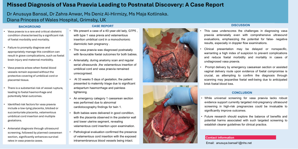
We present a case of a 40 year-old lady, gravida seven para five, with type 1 vasa previa and velamentous insertion umbilical cord in a monochorionic diamniotic twin
pregnancy . The vasa praevia was diagnosed postnatally with favourable foetal outcomes for both babies. Antenatally, during anatomy scan and regular serial
ultrasounds ,the velamentous insertion of umbilical cord and vasa praevia remained unrecognised. At 33 weeks and 5 days of gestation, the patient presented to
maternity triage due to significant antepartum haemorrhage and painless tightening. An emergency category 1 caesarean section was performed due to abnormal
cardiotocography Dndings for twin 1. Both babies were delivered in good condition, with the placenta observed in the posterior wall and lower uterine segment, revealing velamentous cord insertion upon examination. Pathological evaluation confirmed the presence of velamentous cord insertion with the exposed intramembranous blood vessels being intact.
Discussion:
This case underscores the challenges in diagnosing vasa praevia antenatally even with comprehensive ultrasound evaluations, emphasizing the potential for false-
negative results, especially in doppler flow examinations. Clinical presentation may be delayed or nonspeciDc, warranting a high index of suspicion to prevent
complications and reduce foetal morbidity and mortality in cases of undiagnosed vasa praevia. Prompt delivery by emergency caesarean section or assisted vaginal
delivery route upon evidence of foetal compromise is crucial, as attempting to confirm the diagnosis through scanning may jeopardise foetal well-being due to
anticipated brisk foetal blood loss.
Conclusions:
While universal screening for vasa praevia lacks robust evidence support currently, targeted mid-pregnancy ultrasound screening in high-risk pregnancies could be
invaluable to significantly improve outcomes. Future research should explore the balance of benefits and potential harms associated with such targeted screening to
establish clearer guidelines for clinical practice.