2,281 posters, 63 videos, 56 audios, 43 topics, 18 sessions, 6,174 authors, 2,323 institutions
ePostersLive by SciGen Technologies S.A. All rights reserved.
RCOG World Congress 2025
June 23 - 25, 2025 | ExCeL London, UK
EP.1092
Unveiling the Hidden Tumor: A Rare Case of Placental-Chorioangioma Presenting as Polyhydramnios
Part of Topic
High risk pregnancy
Unveiling the Hidden Tumor: A Rare Case of Placental-Chorioangioma Presenting as Polyhydramnios
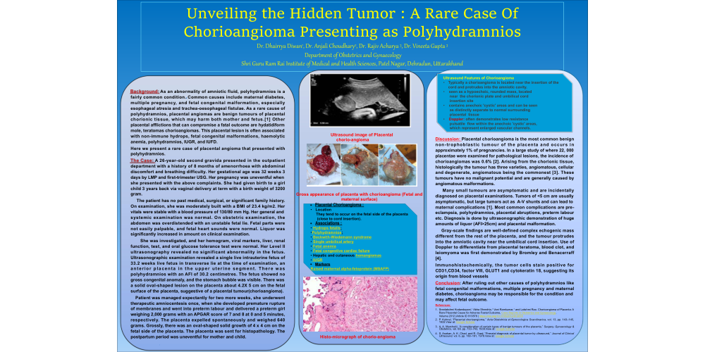
. Background: As an abnormality of amniotic fluid, polyhydramnios is a fairly common condition. Common causes include maternal diabetes, multiple pregnancy, and fetal congenital malformation, especially esophageal atresia and trachea-oesophageal fistulae. As a rare cause of polyhydramnios, placental angiomas are benign tumours of placental chorionic tissue, which may harm both mother and fetus.[1] Other placental afflictions that can compromise a fatal outcome are hydatidiform mole, teratomas chorioangiomas. This placental lesion is often associated with non-immune hydrops, fetal congenital malformations, haemolytic anemia, polyhydramnios, IUGR, and IUFD. Here we present a rare case of placental angioma that presented with polyhydramnios. The Case: A 26-year-old second gravida presented in the outpatient department with a history of 8 months of amenorrhoea with abdominal discomfort and breathing difficulty. Her gestational age was 32 weeks 3 days by LMP and first-trimester USG. Her pregnancy was uneventful when she presented with the above complaints. She had given birth to a girl child 3 years back via vaginal delivery at term with a birth weight of 3200 gram. The patient has no past medical, surgical, or significant family history. On examination, she was moderately built with a BMI of 23.4 kg/m2. Her vitals were stable with a blood pressure of 130/80 mm Hg. Her general and systemic examination was normal. On obstetric examination, the abdomen was overdistended with an unstable fetal lie. Fetal parts were not easily palpable, and fetal heart sounds were normal. Liquor was significantly increased in amount on clinical examination. She was investigated, and her hemogram, viral markers, liver, renal function, test, and oral glucose tolerance test were normal. Her Level II ultrasonography revealed no significant abnormality in the fetus. Ultrasonographic examination revealed a single live intrauterine fetus of 33.2 weeks live fetus in transverse lie at the time of examination, an anterior placenta in the upper uterine segment. There was polyhydramnios with an AFI of 30.2 centimetres. The fetus showed no gross congenital anomaly, and the stomach bubble was visible. There was a solid oval-shaped lesion on the placenta about 4.2X 5 cm on the fetal surface of the placenta, suggestive of a placental tumour(chorioangioma). Patient was managed expectantly for two more weeks, she underwent therapeutic amniocentesis once, when she developed premature rupture of membranes and went into preterm labour and delivered a preterm girl weighing 2,000 grams with an APGAR score of 7 and 8 at 0 and 5 minutes, respectively. The placenta expelled spontaneously and weighed 640 grams. Grossly, there was an oval-shaped solid growth of 4 x 4 cm on the fetal side of the placenta. The placenta was sent for histopathology. The postpartum period was uneventful for mother and child. :
Placental Chorioangioma : Location They tend to occur on the fetal side of the placenta (close to cord insertion). Associations : Hydrops fetalis: Polyhydramnios: Beckwith-Wiedemann syndrome Single umbilical artery Fetal anemia Fetal congestive cardiac failure Hepatic and cutaneous hemangiomas IUGR Markers Raised maternal alpha-fetoprotein (MSAFP)
Ultrasound Features of Chorioangioma Typically a chorioangioma is located near the insertion of the cord and protrudes into the amniotic cavity. seen as a hypoechoic, rounded mass, located near the chorionic plate and umbilical cord insertion site contains anechoic 'cystic' areas and can be seen as distinctly separate to normal surrounding placental tissue Doppler: often demonstrates low resistance pulsatile flow within the anechoic 'cystic' areas, which represent enlarged vascular channels.
Discussion: Placental chorioangioma is the most common benign non-trophoblastic tumour of the placenta and occurs in approximately 1% of pregnancies. In a large study of where 22, 000 placentae were examined for pathological lesions, the incidence of chorioangiomas was 0.6% [2]. Arising from the chorionic tissue, histologically the tumour has three varieties, angiomatous, cellular and degenerate, angiomatous being the commonest [3]. These tumours have no malignant potential and are generally caused by angiomatous malformations. Many small tumours are asymptomatic and are incidentally diagnosed on placental examinations. Tumors of <5 cm are usually asymptomatic, but large tumors act as A-V shunts and can lead to maternal complications [1]. Most common complications are pre-eclampsia, polyhydramnios, placental abruptions, preterm labour etc. Diagnosis is done by ultrasonographic demonstration of huge amounts of liquor (AFI>25cm) and placental malformation. Gray-scale findings are well-defined complex echogenic mass different from the rest of the placenta, and the tumour protrudes into the amniotic cavity near the umbilical cord insertion. Use of Doppler to differentiate from placental teratoma, blood clot, and leiomyoma was first demonstrated by Bromley and Benacerraff [4]. Immunohistochemically, the tumor cells stain positive for CD31,CD34, factor VIII, GLUT1 and cytokeratin 18, suggesting its origin from blood vessels Conclusion: After ruling out other causes of polyhydramnios like fetal congenital malformations, multiple pregnancy and maternal diabetes, chorioangioma may be responsible for the condition and may affect fetal outcome. References: Sreelakshmi Kodandapani,1 Abha Shreshta,1 Vani Ramkumar,1 and Lakshmi Rao. Chorioangioma of Placenta: A Rare Placental Cause for Adverse Foetal Outcome, Case Reports in Obstetrics and Gynaecology. Volume 2012 |Article ID 913878 | https://doi.org/10.1155/2012/913878 P. Kuhnel, “Placental chorioangioma,” Acta Obstetricia et Gynecologica Scandinavica, vol. 13, pp. 143–145, 1933.View at: Google Scholar A. A. Marchetti, “A consideration of certain types of benign tumours of the placenta,” Surgery, Gynaecology & Obstetrics, vol. 68, pp. 733–743, 1939.View at: Google Scholar S. Asokan, A. K. Chad, and R. Gard, “Prenatal diagnosis of placental tumor by ultrasound,” Journal of Clinical Ultrasound, vol. 6, pp. 180–181, 1978.View at: Google Scholar