2,281 posters, 63 videos, 56 audios, 43 topics, 18 sessions, 6,174 authors, 2,323 institutions
ePostersLive by SciGen Technologies S.A. All rights reserved.
RCOG World Congress 2025
June 23 - 25, 2025 | ExCeL London, UK
EP.1058
Cerebral Venous Thrombosis in Pregnancy: A Case Report and Review of Management
Poster Presenter
Authors
Affiliations
Part of Topic
High risk pregnancy
Cerebral Venous Thrombosis (Cin Pregnancy: A Case Report and Review of Management
Author: Shireen Baqir, MBBS, MRCOG Specialty Registrar, James Paget University Hospital
Objective: This report presents a case of CVT in late pregnancy, highlighting the importance of timely diagnosis and effective management. Cerebral venous thrombosis (CVT) is a rare but possibly life-threatening condition, especially during pregnancy. It is often misdiagnosed due to its varied presentation and overlapping symptoms with benign conditions such as migraines and tension headaches.Anticoagulation remains the cornerstone of treatment; however, outcomes may still vary.
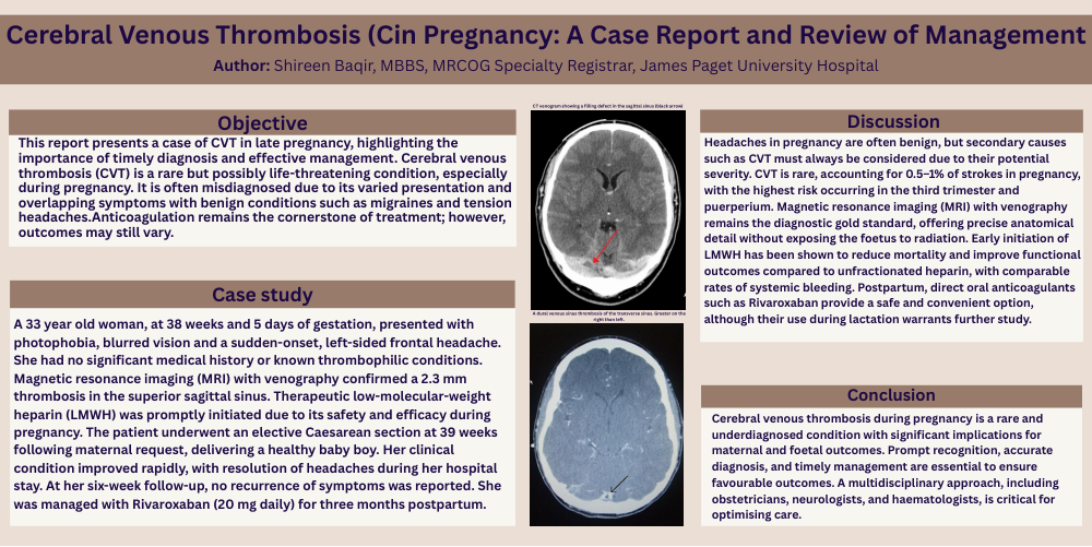
Case study: A 33 year old woman, at 38 weeks and 5 days of gestation, presented with photophobia, blurred vision and a sudden-onset, left-sided frontal headache. She had no significant medical history or known thrombophilic conditions. Magnetic resonance imaging (MRI) with venography confirmed a 2.3 mm thrombosis in the superior sagittal sinus. Therapeutic low-molecular-weight heparin (LMWH) was promptly initiated due to its safety and efficacy during pregnancy. The patient underwent an elective Caesarean section at 39 weeks following maternal request, delivering a healthy baby boy. Her clinical condition improved rapidly, with resolution of headaches during her hospital stay. At her six-week follow-up, no recurrence of symptoms was reported. She was managed with Rivaroxaban (20 mg daily) for three months postpartum
Discussion: Headaches in pregnancy are often benign, but secondary causes such as CVT must always be considered due to their potential severity. CVT is rare, accounting for 0.5–1% of strokes in pregnancy, with the highest risk occurring in the third trimester and puerperium. Magnetic resonance imaging (MRI) with venography remains the diagnostic gold standard, offering precise anatomical detail without exposing the foetus to radiation. Early initiation of LMWH has been shown to reduce mortality and improve functional outcomes compared to unfractionated heparin, with comparable rates of systemic bleeding. Postpartum, direct oral anticoagulants such as Rivaroxaban provide a safe and convenient option, although their use during lactation warrants further study.
Conclusion; Cerebral venous thrombosis during pregnancy is a rare and underdiagnosed condition with significant implications for maternal and foetal outcomes. Prompt recognition, accurate diagnosis, and timely management are essential to ensure favourable outcomes. A multidisciplinary approach, including obstetricians, neurologists, and haematologists, is critical for optimising care.