2,281 posters, 63 videos, 56 audios, 43 topics, 18 sessions, 6,174 authors, 2,323 institutions
ePostersLive by SciGen Technologies S.A. All rights reserved.
RCOG World Congress 2025
June 23 - 25, 2025 | ExCeL London, UK
EP.1022
Management of Placenta Accreta Spectrum: Lessons Learnt over a decade in UK!
Poster Presenter
Authors
Affiliations
Part of Topic
High risk pregnancy
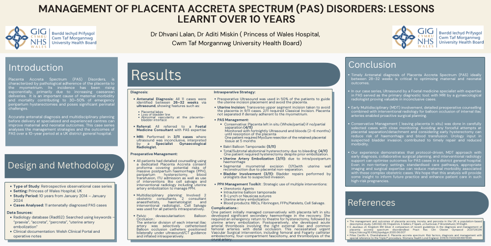
MANAGEMENT OF PLACENTA ACCRETA SPECTRUM (PAS) DISORDERS: LESSONS LEARNT OVER 10 YEARS
Dr Dhvani Lalan, Dr Aditi Miskin ( Princess of Wales Hospital, Cwm Taf Morgannwg University Health Board)
Introduction
Placenta Accreta Spectrum (PAS) Disorders, is characterized by pathological adherence of the placenta to the myometrium. Its incidence has been rising exponentially, primarily due to increasing caesarean deliveries. It is an important cause of maternal morbidity and mortality contributing to 30–50% of emergency peripartum hysterectomies and poses significant perinatal challenges.
Accurate antenatal diagnosis and multidisciplinary planning before delivery at specialized and experienced centres can improve maternal and neonatal outcomes. This case series analyses the management strategies and the outcomes of PAS over a 10-year period at a UK district general hospital.
Diagnosis:
Antenatal Diagnosis: All 11 cases were identified between 26–32 weeks via ultrasound, showing features such as:
Placental lakes
Loss of bladder line
Abnormal vascularity at the placenta– bladder interface
Intraoperative Strategy:
Preoperative Ultrasound was used in 50% of the patients to guide the uterine incision placement and avoid the placenta.
Uterine Incision: Transverse upper segment incision taken to avoid the placenta in 9/11 cases. 2/11 required Classical Incision. Placenta not separated if densely adherent to the myometrium.
PAS Management:
Referral: All referred to a Foetal Medicine Consultant with PAS expertise
Conservative: Placenta left in situ (Whole/partial) if no/partial separation (4/11)
Monitored with fortnightly Ultrasound and bloods (2–6 months) until resorption of the placenta
.MRI: Performed in 3/11 cases where ultrasound was inconclusive, interpreted by a Specialist Gynaecological Radiologist.
One patient required MyoSure resection of the retained placental tissue at 5 months
Preoperative Management:
All patients had detailed counselling using a dedicated Placenta Accreta consent proforma covering potential risks of massive postpartum haemorrhage (PPH), peripartum hysterectomy, blood transfusion, ITU admission as well as use of interventions like cell salvage and interventional radiology including uterine artery embolization to manage PPH.
Multidisciplinary planning: Involved 2 obstetric consultants, 2 consultant anaesthetists, haematologist and interventional radiologist.. Cell Salvage was used for all patients intraoperatively.
Pelvic devascularisation: Balloon Occlusion :
The anterior division of each internal iliac artery was selectively catheterised Balloon occlusion catheters positioned bilaterally under ultrasound/CT guidance and inflated intraoperatively.
Bakri Balloon Tamponade: (5/11)
Total/Subtotal abdominal hysterectomy due to bleeding: (4/11)
One patient required hysterectomy despite prior embolization.
Uterine Artery Embolization (3/11): due to intra/postpartum haemorrhage
Segmental myometrial excision (1/11)with uterine wall reconstruction due to placental non-separation.
Bladder Involvement (3/11): Bladder repairs performed by urologists due to suspected invasion.
PPH Management Toolkit : Strategic use of multiple interventions:
Uterotonic Agents
Intrauterine balloon tamponade
B-Lynch or Nausicaa sutures
Uterine artery embolization
Blood products: RBCs, Fibrinogen, FFPs,Platelets, Cell Salvage.
Complications :
One patient initially managed conservatively with placenta left in situ developed significant secondary haemorrhage in the recovery. She required an emergency return to theatre for hysterectomy, followed by uterine artery embolization. Postoperatively, she developed acute arterial thrombosis involving the right external iliac and common femoral arteries with distal occlusion. This necessitated urgent Vascular Surgical intervention, including femoral and Fogarty catheter embolectomy, four-compartment fasciotomy, and thrombolysis of the crural artery.
Conclusion
Timely Antenatal diagnosis of Placenta Accreta Spectrum (PAS) ideally between 28–32 weeks is critical to optimising maternal and neonatal outcomes.
In our case series, Ultrasound by a Foetal medicine specialist with expertise in PAS served as the primary diagnostic tool; with MRI by a gynaecological radiologist proving valuable in inconclusive cases.
Early Multidisciplinary (MDT) involvement, detailed preoperative counselling combined with interventional radiology for balloon occlusion of internal iliac arteries enabled proactive surgical planning.
Conservative Management ( leaving placenta in situ) was done in carefully selected cases with close monitoring. Avoiding any forceful attempts at placental separation/detachment and considering early hysterectomy can reduce risk of haemorrhage and blood transfusion. Urology input in suspected bladder invasion, contributed to timely repair and reduced morbidity.
Our experience demonstrates that protocol-driven, MDT approach with early diagnosis, collaborative surgical planning, and interventional radiology support can optimise outcomes for PAS cases in a district general hospital. Even in non-tertiary settings, standardised care pathways, appropriate imaging and surgical expertise can reduce maternal morbidity associated with these complex obstetric cases. We hope that this analysis will provide some insight to inform future practice and enhance patient care in such high-risk pregnancies.
Results
Design and Methodology
Type of Study: Retrospective observational case series Setting: Princess of Wales Hospital, UK
Study Period: 10 years from January 2014 – January 2024
Cases Analysed: 11 antenatally diagnosed PAS cases
Data Sources:
Radiology database (RadIS2): Searched using keywords – “praevia”, “accreta”, “percreta”, “uterine artery embolization”
Clinical documentation: Welsh Clinical Portal and operative notes
References
The management and outcomes of placenta accreta, increta, and percreta in the UK: a population-based descriptive study. (UKOSS)-KE Fitzpatrick, S Sellers, P Spark, JJ Kurinczuk, P Brocklehurst, M Knight.
E Jauniaux JC Kingdom RM Silver A comparison of recent guidelines in the diagnosis and management of placenta accreta spectrum disordersBest Pract Res Clin Obstet Gynaecol 20217210216 https://doi.org/10.1016/j.bpobgyn.2020.06.007
Piñas Carrillo A, Chandraharan E. Placenta accreta spectrum: Risk factors, diagnosis and management with special reference to the Triple P procedure. Womens Health Lond England. 2019;15:1745506519878081.